Recommended Products
biological source
rabbit
Quality Level
100
500
conjugate
unconjugated
antibody form
Ig fraction of antiserum
antibody product type
primary antibodies
clone
polyclonal
description
For In Vitro Diagnostic Use in Select Regions (See Chart)
form
buffered aqueous solution
species reactivity
human
packaging
vial of 0.1 mL concentrate (393A-14)
vial of 0.5 mL concentrate (393A-15)
bottle of 1.0 mL predilute (393A-17)
vial of 1.0 mL concentrate (393A-16)
bottle of 7.0 mL predilute (393A-18)
manufacturer/tradename
Cell Marque™
technique(s)
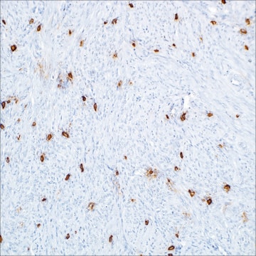
immunohistochemistry (formalin-fixed, paraffin-embedded sections): 1:25-1:100
control
sebaceous neoplasms (intracytoplasmic lipid droplet)
shipped in
wet ice
storage temp.
2-8°C
visualization
membranous
Gene Information
human ... PLIN2(123)
General description
Of 25 sebaceous carcinomas, 23 (92%) were also labeled with a similar pattern. Additionally, in cases of poorly differentiated sebaceous carcinoma (n=11), in which sebaceous differentiation could not have been reliably interpreted in H&E sections, adipophilin highlighted the sebocytes with a strong membranous labeling of intracytoplasmic lipid droplets in 9 of 11 cases (82%). Moreover, 10 of 10 (100%) xanthelasmas, 9 of 10 (90%) xanthogranulomas, 6 of 6 (100%) xanthomas, and 9 of 13 (63%) metastatic renal cell carcinomas were also weakly-to- moderately positive for adipophilin. Expression of adipophilin with a membranous pattern of staining was not seen in any of the other clear cell lesions of the skin, including basal and squamous cell carcinomas, trichilemmomas, clear cell hidradenomas, or balloon cell nevi. Interestingly, a nonspecific granular uptake of anti-adipophilin was seen in adjacent macrophages, keratohyalin granules of epithelial squamous cells, and some tumor cells. Therefore, this anti-adipophilin is suitable for immunostaining formalin-fixed, paraffin-embedded tissue and is helpful in the identification of intracytoplasmic lipids, as seen in sebaceous lesions. It is especially helpful in identifying intracytoplasmic lipid vesicles in poorly differentiated sebaceous carcinomas in challenging cases such as small periocular biopsy specimens.
Quality
 IVD |  IVD |  IVD |  RUO |
Linkage
Physical form
Preparation Note
Other Notes
Legal Information
Not finding the right product?
Try our Product Selector Tool.
Certificates of Analysis (COA)
Search for Certificates of Analysis (COA) by entering the products Lot/Batch Number. Lot and Batch Numbers can be found on a product’s label following the words ‘Lot’ or ‘Batch’.
Already Own This Product?
Find documentation for the products that you have recently purchased in the Document Library.
Articles
IHC antibodies enhance dermatopathology beyond H&E stained slides, improving techniques and applications for dermatological research.
IHC antibodies enhance dermatopathology beyond H&E stained slides, improving techniques and applications for dermatological research.
IHC antibodies enhance dermatopathology beyond H&E stained slides, improving techniques and applications for dermatological research.
IHC antibodies enhance dermatopathology beyond H&E stained slides, improving techniques and applications for dermatological research.
Our team of scientists has experience in all areas of research including Life Science, Material Science, Chemical Synthesis, Chromatography, Analytical and many others.
Contact Technical Service


