Skip to Content
Merck
All Photos(2)

Documents

MABC604

Sigma-Aldrich

Anti-MLKL Antibody, clone 3H1

clone 3H1, from rat

Synonym(s):

Mixed lineage kinase domain-like protein, MLKL

Sign Into View Organizational & Contract Pricing


About This Item

UNSPSC Code:
12352203
eCl@ss:
32160702
NACRES:
NA.41

biological source

rat

Quality Level

antibody form

purified antibody

antibody product type

primary antibodies

clone

3H1, monoclonal

species reactivity

mouse

technique(s)

immunocytochemistry: suitable
immunoprecipitation (IP): suitable
western blot: suitable

isotype

IgG

NCBI accession no.

UniProt accession no.

shipped in

wet ice

target post-translational modification

unmodified

Gene Information

mouse ... Mlkl(74568)

General description

MLKL, also known as Mixed lineage kinase domain-like protein, and encoded by the gene MLKL, is an interesting protein that is required for the execution of programmed necrosis or necroptosis, for example in response to TNF-alpha induced necrosis. MLKL is a component of the "necrosome," the multiprotein complex that triggers tumor necrosis factor (TNF)-induced cell death by necroptosis. The programmed necrosis induced by TNF-α also requires the activities of the receptor-interacting serine-threonine kinases RIP1 and RIP3 and they too interact directly with the mixed lineage kinase domain-like protein MLKL. RIP3 is an essential upstream kinase in necroptosis. The pseudokinase MLKL functions as a substrate of RIP3 to mediate downstream signaling and when complexed to MLKL the RIP3-MLKL complex is stabilized by AMP-PNP to adopt an inactive conformation.

Immunogen

Epitope: Brace region.
KLH-conjugated linear peptide corresponding to a sequence from the two-helix linker (Brace) region N-terminal to the pseudokinase domain of mouse MLKL (Hildebrand, J.M., et al. (2014). Proc. Natl. Acad. Sci. U.S.A. 111(42):15072-15077; Murphy, J.M., et al. (2013). Immunity. 39(3):443-453).

Application

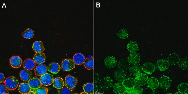
Immunocytochemistry Analysis: A representative lot detected MLKL in mouse dermal fibroblasts (MDFs) from wild-type, but not Mlkl-/- mice (Courtesy of Prof. James M. Murphy, Walter and Eliza Hall Institute, Australia).

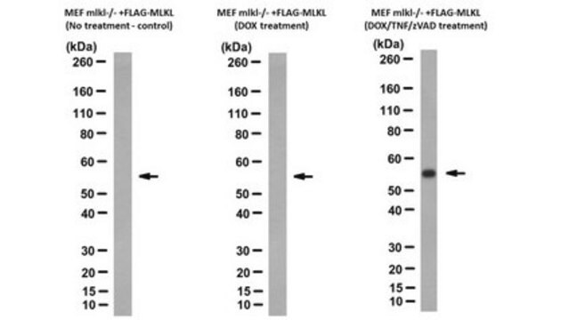
Immunoprecipitation Analysis: A representative lot immunoprecipitated MLKL from soluble extract of Mlkl-/- mouse dermal fibroblasts (MDFs) harboring doxycycline-inducible wild-type mouse MLKL expression construct only after, but not before doxycycline treatment (Murphy, J.M., et al. (2013). Immunity. 39(3):443-453).

Western Blotting Analysis: A representative lot detected Q-VD-OPh (TSQ; Cat. No. 551476) treatment-induced membrane translocation of murine and equine MLKL N-terminal fragment (a.a. 1-180 and 1-189, respectively) exogenously expressed in mouse dermal fibroblasts (MDFs) from Mlkl-/- mice (Tanzer, M.C., et al. (2016). Cell Death Differ.. In press).

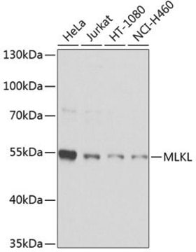
Western Blotting Analysis: A representative lot detected MLKL in human HT-29 and U937 cells (Tanzer, M.C., et al. (2015). Biochem. J. 471(2):255-265).

Western Blotting Analysis: Representative lots detected MLKL membrane translocation in mouse dermal fibroblasts (MDFs) upon Q-VD-OPh (TSQ; Cat. No. 551476) treatment (Tanzer, M.C., et al. (2015). Biochem. J. 471(2):255-265; Hildebrand, J.M., et al. (2014). Proc. Natl. Acad. Sci. U.S.A. 111(42):15072-15077).

Western Blotting Analysis: Representative lots detected MLKL expression in L292 mouse fibroblasts, as well as in mouse dermal fibroblasts (MDFs), mouse embryonic fibroblasts (MEFs) and bone marrow derived macrophages (BMDMs) from wild-type, but not Mlkl-/- mice (Cook, W.D., et al. (2014). Cell Death Differ. 21(10):1600-1612; Murphy, J.M., et al. (2013). Immunity. 39(3):443-453).

Western Blotting Analysis: A representative lot detected recombinant full-length mouse MLKL as well as a.a. 1-180 and a.a. 124-464, but not a.a. 1-125 or a.a. 179-464, MLKL fragments (Hildebrand, J.M., et al. (2014). Proc. Natl. Acad. Sci. U.S.A. 111(42):15072-15077).

Western Blotting Analysis: A representative lot detected MLKL expression in all tissues tested except brain from wild-type mice, while no target band was seen in any tissues from Mlkl-/- mice (Murphy, J.M., et al. (2013). Immunity. 39(3):443-453).
This Anti-MLKL Antibody, clone 3H1 is validated for use in Immunocytochemistry, Immunoprecipitation, and Western Blotting for the detection of MLKL.

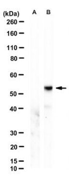
Quality

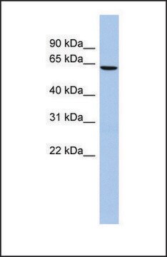
Evaluated by Western Blotting in mouse heart tissue lysate.

Western Blotting Analysis: 0.5 µg/mL of this antibody detected MLKL in 10 µg of mouse heart tissue lysate.

Target description

~52 kDa observed. 54.48/30.28 (human isoform 1/2) and 54.32/53.38 kDa (mouse isoform 1/2) calculated. Uncharacterized band(s) may be observed in some cell lysates.

Physical form

Format: Purified
Purified rat IgG in buffer containing 0.1 M Tris-Glycine (pH 7.4), 150 mM NaCl with 0.05% sodium azide.

Other Notes

Concentration: Please refer to the Certificate of Analysis for the lot-specific concentration.

Not finding the right product?  

Try our Product Selector Tool.

Storage Class Code

12 - Non Combustible Liquids

WGK

WGK 1

Flash Point(F)

Not applicable

Flash Point(C)

Not applicable


Certificates of Analysis (COA)

Search for Certificates of Analysis (COA) by entering the products Lot/Batch Number. Lot and Batch Numbers can be found on a product’s label following the words ‘Lot’ or ‘Batch’.

Already Own This Product?

Find documentation for the products that you have recently purchased in the Document Library.

Visit the Document Library

RIPK1- and RIPK3-induced cell death mode is determined by target availability.
Cook, WD; Moujalled, DM; Ralph, TJ; Lock, P; Young, SN; Murphy, JM; Vaux, DL
Cell Death and Differentiation null
D M Moujalled et al.
Cell death & disease, 5, e1086-e1086 (2014-03-01)
Necroptosis is a mechanism by which cells can kill themselves that does not require caspase activity or the presence of the pro-apoptotic Bcl-2 family members Bax or Bak. It has been reported that RIPK3 (receptor interacting protein kinase 3) activates
A V Jacobsen et al.
Cell death & disease, 7, e2051-e2051 (2016-01-19)
Necroptosis is a caspase-independent form of regulated cell death that has been implicated in the development of a range of inflammatory, autoimmune and neurodegenerative diseases. The pseudokinase, Mixed Lineage Kinase Domain-Like (MLKL), is the most terminal known obligatory effector in
M C Tanzer et al.
Cell death and differentiation, 23(7), 1185-1197 (2016-02-13)
The pseudokinase, MLKL (mixed-lineage kinase domain-like), is the most terminal obligatory component of the necroptosis cell death pathway known. Phosphorylation of the MLKL pseudokinase domain by the protein kinase, receptor interacting protein kinase-3 (RIPK3), is known to be the key
Sefi Zargarian et al.
PLoS biology, 15(6), e2002711-e2002711 (2017-06-27)
Necroptosis is a regulated, nonapoptotic form of cell death initiated by receptor-interacting protein kinase-3 (RIPK3) and mixed lineage kinase domain-like (MLKL) proteins. It is considered to be a form of regulated necrosis, and, by lacking the "find me" and "eat

Our team of scientists has experience in all areas of research including Life Science, Material Science, Chemical Synthesis, Chromatography, Analytical and many others.

Contact Technical Service