Skip to Content
Merck
All Photos(2)

Documents

04-147

Sigma-Aldrich

Anti-ASC Antibody, clone 2EI-7

clone 2EI-7, from mouse

Synonym(s):

Caspase recruitment domain-containing protein 5, PYD and CARD domain containing, PYD and CARD domain-containing protein, Target of methylation-induced silencing 1, apoptosis-associated speck-like protein containing a CARD, caspase recruitment domain prot

Sign Into View Organizational & Contract Pricing


About This Item

UNSPSC Code:
12352203
eCl@ss:
32160702
NACRES:
NA.41

biological source

mouse

Quality Level

antibody form

purified antibody

antibody product type

primary antibodies

clone

2EI-7, monoclonal

species reactivity

mouse

species reactivity (predicted by homology)

human (based on 100% sequence homology)

technique(s)

flow cytometry: suitable
western blot: suitable

isotype

IgG1κ

NCBI accession no.

UniProt accession no.

shipped in

wet ice

target post-translational modification

unmodified

Gene Information

human ... PYCARD(29108)

General description

Apoptosis is regulated by death domain (DD) and/or caspase recruitment domain (CARD) containing molecules and a caspase family of proteases. CARD containing cell death regulators include RAIDD, RICK, Bcl10, Apaf-1, ARC, caspase-2 and caspase-9. A novel CARD domain containing protein has been identified in human and mouse and designated ASC and TMS1. Irregular expression of ASC/TMS1 induced apoptosis through activation of caspase-9 and inhibited the survival of human breast cancer cells. Overexpression of ASC/TMS1 induced DNA fragmentation. ASC/TMS1 is expressed in a variety of human and mouse tissues.

Specificity

This antibody recognizes ASC.

Immunogen

Epitope: Unknown
Full length recombinant human ASC.

Application

Detect ASC using this Anti-ASC Antibody, clone 2EI-7 validated for use in WB, FC.
Research Category
Inflammation & Immunology
Research Sub Category
Apoptosis - Additional

Quality

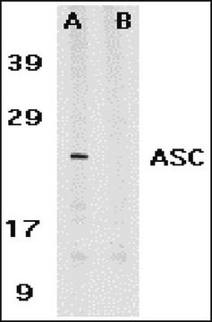
Evaluated by Western Blot on mouse spleen tissue lysate.

Western Blot Analysis: 0.5 µg/mL of this antibody detected ASC in mouse spleen tissue lysate.

Target description

~22 kDa

Physical form

Format: Purified
Protein G Purified
Purified mouse monoclonal IgG1κ in buffer containing 0.1 M Tris-Glycine (pH 7.4, 150 mM NaCl) with 0.05% sodium azide.

Storage and Stability

Stable for 1 year at 2-8°C from date of receipt.

Analysis Note

Control
Mouse spleen tissue lysate

Other Notes

Concentration: Please refer to the Certificate of Analysis for the lot-specific concentration.

Disclaimer

Unless otherwise stated in our catalog or other company documentation accompanying the product(s), our products are intended for research use only and are not to be used for any other purpose, which includes but is not limited to, unauthorized commercial uses, in vitro diagnostic uses, ex vivo or in vivo therapeutic uses or any type of consumption or application to humans or animals.

Not finding the right product?  

Try our Product Selector Tool.

Storage Class Code

12 - Non Combustible Liquids

WGK

WGK 1

Flash Point(F)

Not applicable

Flash Point(C)

Not applicable


Certificates of Analysis (COA)

Search for Certificates of Analysis (COA) by entering the products Lot/Batch Number. Lot and Batch Numbers can be found on a product’s label following the words ‘Lot’ or ‘Batch’.

Already Own This Product?

Find documentation for the products that you have recently purchased in the Document Library.

Visit the Document Library

Sonal Khare et al.
Nature immunology, 15(4), 343-353 (2014-02-18)
The innate immune system responds to infection and tissue damage by activating cytosolic sensory complexes called 'inflammasomes'. Cytosolic DNA is sensed by AIM2-like receptors (ALRs) during bacterial and viral infections and in autoimmune diseases. Subsequently, recruitment of the inflammasome adaptor
Wei Zhu et al.
Neuroscience, 343, 77-84 (2016-12-08)
Lipopolysaccharide (LPS) might affect the central nervous system by causing neuroinflammation, which subsequently leads to brain damage and dysfunction. In this study, we evaluated the role of nod-like receptor pyrin domain-containing protein 3 (NLRP3) inflammasome activation in long-term behavioral alterations
Tiantian Tang et al.
Nature communications, 8(1), 202-202 (2017-08-06)
The NLRP3 inflammasome can sense different pathogens or danger signals, and has been reported to be involved in the development of many human diseases. Potassium efflux and mitochondrial damage are both reported to mediate NLRP3 inflammasome activation, but the underlying
Sung-Soo Jung et al.
American journal of physiology. Lung cellular and molecular physiology, 308(10), L1058-L1067 (2015-03-15)
Inflammasomes are cytosolic protein complexes that promote the cleavage of caspase-1, which leads to the maturation and secretion of proinflammatory cytokines, including interleukin-1β (IL-1β) and IL-18. Among the known inflammasomes, the nucleotide-binding domain, leucine-rich-containing family, pyrin domain-containing-3 (NLRP3)-dependent inflammasome is
Teneema Kuriakose et al.
Journal of immunology (Baltimore, Md. : 1950), 200(4), 1489-1495 (2018-01-13)
Innate immune sensing of influenza A virus (IAV) induces activation of various immune effector mechanisms, including the nucleotide and oligomerization domain, leucine-rich repeat-containing protein family, pyrin domain containing 3 (NLRP3) inflammasome and programmed cell death pathways. Although type I IFNs

Our team of scientists has experience in all areas of research including Life Science, Material Science, Chemical Synthesis, Chromatography, Analytical and many others.

Contact Technical Service