Skip to Content
Merck
All Photos(2)

Documents

MABS186

Sigma-Aldrich

Anti-LOX-1, clone 9E12.1

clone 9E12.1, from mouse

Synonym(s):

LOX-1, Oxidized low-density lipoprotein receptor 1, Ox-LDL receptor 1, C-type lectin domain family 8 member A, Lectin-like oxidized LDL receptor 1, Lectin-like oxLDL receptor 1, hLOX-1, Lectin-type oxidized LDL receptor 1, Oxidized low-density lipoprotei

Sign Into View Organizational & Contract Pricing


About This Item

UNSPSC Code:
12352203
eCl@ss:
32160702
NACRES:
NA.41

biological source

mouse

Quality Level

antibody form

purified immunoglobulin

antibody product type

primary antibodies

clone

9E12.1, monoclonal

species reactivity

human

technique(s)

immunohistochemistry: suitable (paraffin)
western blot: suitable

isotype

IgG2aκ

NCBI accession no.

UniProt accession no.

shipped in

wet ice

target post-translational modification

unmodified

Gene Information

human ... OLR1(4973)

General description

LOX1 is a plasma membrane receptor, that also has a free soluble receptor form, that is the receptor that mediates the recognition, internalization and degradation of oxidatively modified low density lipoprotein (oxLDL) by vascular endothelial cells. OxLDL is a marker of atherosclerosis that induces vascular endothelial cell activation and dysfunction, resulting in pro-inflammatory responses, pro-oxidative conditions and apoptosis. Its association with oxLDL induces the activation of NF-kappa-B through an increased production of intracellular reactive oxygen and a variety of pro-atherogenic cellular responses including a reduction of nitric oxide (NO) release, monocyte adhesion and apoptosis. LOX1 is also the receptor for HSP70, and LOX1 acts as a leukocyte adhesion molecule on glycosylated proteins present on platelets, monocytes, bacteria, apoptotic cells in inflammatory responses.

Immunogen

GST-tagged recombinant protein corresponding to human LOX-1.

Application

Anti-LOX-1 Antibody, clone 9E12.1 is an antibody against LOX-1 for use in Immunohistochemistry and Immunocytochemistry (Paraffin).
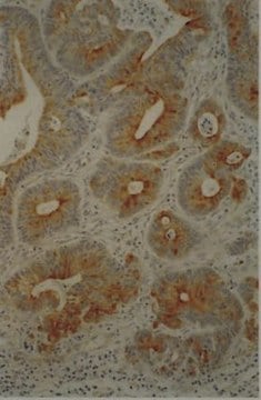
Immunohistochemistry Analysis: A 1:50 dilution from a representative lot detected LOX-1 in human placenta tissue.
Research Category
Signaling
Research Sub Category
Signaling Neuroscience

Quality

Evaluated by Western Blotting in KG-1 cell lysate.

Western Blotting Analysis: A 1:500 dilution of this antibody detected LOX-1 in 200 µg of KG-1 cell lysate.

Target description

~35 kDa observed. The calculated molecular weight of this protein is 31 kDa, but can be observed at ~33-37 kDa due to high glycosylation.

Physical form

Format: Purified
Protein G Purified
Purified mouse monoclonal IgG2aκ in buffer containing 0.1 M Tris-Glycine (pH 7.4), 150 mM NaCl with 0.05% sodium azide.

Storage and Stability

Stable for 1 year at 2-8°C from date of receipt.

Other Notes

Concentration: Please refer to lot specific datasheet.

Disclaimer

Unless otherwise stated in our catalog or other company documentation accompanying the product(s), our products are intended for research use only and are not to be used for any other purpose, which includes but is not limited to, unauthorized commercial uses, in vitro diagnostic uses, ex vivo or in vivo therapeutic uses or any type of consumption or application to humans or animals.

Not finding the right product?  

Try our Product Selector Tool.

Storage Class Code

12 - Non Combustible Liquids

WGK

WGK 1

Flash Point(F)

Not applicable

Flash Point(C)

Not applicable


Certificates of Analysis (COA)

Search for Certificates of Analysis (COA) by entering the products Lot/Batch Number. Lot and Batch Numbers can be found on a product’s label following the words ‘Lot’ or ‘Batch’.

Already Own This Product?

Find documentation for the products that you have recently purchased in the Document Library.

Visit the Document Library

Ayumi Hashimoto et al.
Clinical cancer research : an official journal of the American Association for Cancer Research, 27(21), 5961-5978 (2021-08-20)
To evaluate the mechanisms of how therapeutic upregulation of the transcription factor, CCAAT/enhancer-binding protein alpha (C/EBPα), prevents tumor progression in patients with advanced hepatocellular carcinoma (HCC) and in different mouse tumor models. We conducted a phase I trial in 36

Our team of scientists has experience in all areas of research including Life Science, Material Science, Chemical Synthesis, Chromatography, Analytical and many others.

Contact Technical Service