Skip to Content
Merck

Skip To

MABC1640

Anti-RIPK3/RIP3 Antibody, clone 1H2

Sign In to View Organizational & Contract Pricing.

Select a Size

Change View
Size/SKUAvailabilityPrice
100 μL

Available to ship TODAYfromAreál Kühne+Nagel spol. s r.o.

11 000,00 CZK

About This Item

NACRES:
NA.41
UNSPSC Code:
12352203
Conjugate:
unconjugated
Clone:
1H2, monoclonal
Application:
IF, WB
Citations:
-

11 000,00 CZK


Available to ship TODAYDetails


​
Request a Custom Order
Technical Service
Need help? Our team of experienced scientists is here for you.
Let Us Assist


biological source

rat

Quality Segment

conjugate

unconjugated

antibody form

purified antibody

antibody product type

primary antibodies

clone

1H2, monoclonal

mol wt

calculated mol wt 56.89 kDa, observed mol wt ~57 kDa

species reactivity

human

packaging

antibody small pack of 100 μL

technique(s)

immunofluorescence: suitable, western blot: suitable

isotype

IgG2aκ

UniProt accession no.

shipped in

dry ice

storage temp.

2-8°C

target post-translational modification

unmodified

General description

Receptor-interacting serine/threonine-protein kinase 3 (UniProt: Q9Y572; also known as EC:2.7.11.1, RIP-like protein kinase 3, Receptor-interacting protein 3, RIP-3) is encoded by the RIPK3 (also known as RIP3) gene (Gene ID: 11035) in human. RIPK3 is a serine/threonine protein kinase that is expressed in embryo and adult spleen, liver, testis, brain, and lung. Its protein kinase domain is localized in amino acids 21 to 287 in the N-terminal half and it also has a C-terminal RIP homolytic interaction motif (RHIM; aa 450-466), which mediates interaction with the RHIM motif of RIPK1 and facilitate the formation of a necrosome. RIPK3 is shown to activate both necroptosis and apoptosis. Activated RIPK3 forms a necrosis-inducing complex and mediates phosphorylation of MLKL, promoting MLKL localization to the plasma membrane and execution of programmed necrosis that is characterized by calcium influx and plasma membrane damage. Its location is mainly cytoplasmic, however, following infection, it can also be detected in the nucleus. RIPK3 also regulates apoptosis that is dependent on RIPK1, FADD, and CASP8, and is independent of MLKL and RIPK3 kinase activity. It phosphorylates RIPK1 and then RIPK1 and RIPK3 undergo reciprocal auto- and trans-phosphorylation. In some cell types, RIPK3 is also reported to restrict viral replication by promoting cell death-independent responses. Clone 8G7 is raised against an extended kinase domain (residues 2-353) of mouse RIPK3 and can detect forms that may not be detected by antibodies directed towards the C-terminus of the full-length RIPK3. (Ref.: Samson, AL., et al. (2021). Cell Death Differ. https://doi.org/10.1038/s41418-021-00742-x; Petrie, EJ., et al. (2019). Cell Rep. 28(13); 3309-3319).

Immunogen

Recombinant fragment corresponding to 355 amino acids from the extended kinase domain of human Receptor-interacting serine/threonine-protein kinase 3 (RIPK3).

Application

Quality Control Testing

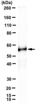
Evaluated by Western Blotting in HT-29 cell lysate.

Western Blotting Analysis: A 1:500 dilution of this antibody detected RIPK3/RIP3 in HT-29 cell lysate.

Tested Applications

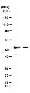
Western Blotting Analysis: A representative lot detected RIPK3/RIP3 in Western Blotting applications (Samson, A.L., et. al. (2020). Nat Commun. 11(1):3151).

Immunofluorescence Analysis: A representative lot detected RIPK3/RIP3 in Immunofluorescence applications (Samson, A.L., et. al. (2021). Cell Death Differ. https://doi.org/10.1038/s41418-021-00742-x).

Note: Actual optimal working dilutions must be determined by end user as specimens, and experimental conditions may vary with the end user
Anti-RIPK3/RIP3, clone 1H2, Cat. No. MABC1640, is a rat monoclonal antibody that detects RIPK3/RIP3 and is tested for use in Immunofluorescence and Western Blotting.

Biochem/physiol Actions

Clone 1H2 is a rat monoclonal antibody that detects human Receptor-interacting serine/threonine-protein kinase (RIP3). It targets an epitope within the extended kinase domain that includes most of the N-terminal half and a part of C-terminal half.

Physical form

Purified rat monoclonal antibody IgG2a in buffer containing 0.1 M Tris-Glycine (pH 7.4), 150 mM NaCl with 0.05% sodium azide.

Preparation Note

Recommend storage at +2°C to +8°C. For long term storage antibodies can be kept at -20°C. Avoid repeated freeze-thaws.

Other Notes

Concentration: Please refer to the Certificate of Analysis for the lot-specific concentration.

Disclaimer

Unless otherwise stated in our catalog or other company documentation accompanying the product(s), our products are intended for research use only and are not to be used for any other purpose, which includes but is not limited to, unauthorized commercial uses, in vitro diagnostic uses, ex vivo or in vivo therapeutic uses or any type of consumption or application to humans or animals.

Compare Similar Items

View Full Comparison

Show Differences

1 of 1

This Item
MABC1595HPA055087SAB2103903
description

-

description

-

description

Prestige Antibodies® Powered by Atlas Antibodies, affinity isolated antibody, buffered aqueous glycerol solution

description

affinity isolated antibody

conjugate

unconjugated

conjugate

unconjugated

conjugate

unconjugated

conjugate

unconjugated

antibody form

purified antibody

antibody form

purified antibody

antibody form

affinity isolated antibody

antibody form

affinity isolated antibody

species reactivity

human

species reactivity

mouse

species reactivity

human

species reactivity

human

biological source

rat

biological source

rat

biological source

rabbit

biological source

rabbit

clone

1H2, monoclonal

clone

8G7, monoclonal

clone

polyclonal

clone

polyclonal

mol wt

observed mol wt ~57 kDa, calculated mol wt 56.89 kDa

mol wt

calculated mol wt 53.32 kDa, observed mol wt ~53 kDa

mol wt

-

mol wt

57 kDa


Still not finding the right product?

Try our Product Selector Tool to narrow your options


Storage Class

12 - Non Combustible Liquids

WGK

WGK 1

Flash Point (°F)

Not applicable

Flash Point (°C)

Not applicable


Choose from one of the most recent versions:

Certificates of Analysis (COA)

Lot/Batch Number

It looks like we've run into a problem, but you can still download Certificates of Analysis from our Documents section.

If you need assistance, please contact Customer Support

Already Own This Product?

Find documentation for the products that you have recently purchased in the Document Library.

Visit the Document Library


Questions

Reviews

No rating value

Active Filters