Skip to Content
Merck
All Photos(4)

Documents

AB1928

Sigma-Aldrich

Anti-Integrin α5 Antibody, CT, Intracellular

serum, Chemicon®

Synonym(s):

CD49e, Fibronectin Receptor alpha Subunit

Sign Into View Organizational & Contract Pricing


About This Item

UNSPSC Code:
12352203
eCl@ss:
32160702
NACRES:
NA.41

biological source

rabbit

Quality Level

antibody form

serum

antibody product type

primary antibodies

clone

polyclonal

species reactivity

human, chicken, hamster, rat, mouse

manufacturer/tradename

Chemicon®

technique(s)

ELISA: suitable
immunocytochemistry: suitable
immunofluorescence: suitable
immunohistochemistry: suitable
immunoprecipitation (IP): suitable
radioimmunoassay: suitable
western blot: suitable

NCBI accession no.

UniProt accession no.

shipped in

dry ice

target post-translational modification

unmodified

Gene Information

human ... ITGA5(3678)

General description

Integrins are a family of dimeric, transmembrane proteins that mediate cell-cell and extracellular matrix adhesion. Signals transduced by integrins play a role in many biological processes, including cell growth, differentiation, migration and apoptosis. The integrin family is composed of at least 15 alpha and 8 beta subunits that may form over twenty different alpha-beta non-covalently bound dimeric combinations on the cell surface. The alpha subunits all have some homology to each other, as do the beta subunits. Both of the subunits contribute to the binding of the ligand. Integrin alpha subunits contain seven weak sequence repeats in the N-terminal region, which may be important in ligand binding, and have been predicted to fold cooperatively into a single beta-propeller domain with seven beta-sheets. The alpha-3 subunit (CD49c) is highly concentrated in epithelial cells where it strongly adheres to Laminin-5 and Laminin-5 induced rapid adhesion can be blocked by antibodies against the alpha-3 integrin subunit. The alpha-3 subunit exists in two different splice variants, denoted as "A" and "B". The only difference that results from this differential splicing is a total change in the cytoplasmic domain, while the extracellular domain stays the same. Knock-out mice lacking this subunit show prenatal lethality and abnormalities in the kidneys.

Specificity

Antibody recognizes integrin alpha5. Specificity determined by immunoprecipitation from a detergent extract of 35S methionine labeled endothelial cells. No cross-reactivity to alpha1, alpha2, alpha3, alpha4, alpha6 or alphaV. The antibody recognizes an epitope on the cytoplasmic domain of the protein, and it is thus, intracellular.
Reactivity with other species has not been determined.

Immunogen

Synthetic peptide derived from the C-terminal sequence (cytoplasmic domain) of the human alpha5 integrin subunit.

Application

Anti-Integrin α5 Antibody, C-terminus, Intracellular detects level of Integrin α5 & has been published & validated for use in ELISA, IC, IF, IH, IP, RIA & WB.
Western Blot Analysis:
1:1000 dilution of a previous lot was used.

ELISA:
1:500-1:2000 dilution of a previous lot was used in ELISA.

Immunoprecipitation:
Recommended use of 5 μL of antibody for 5x10⁶ cells.

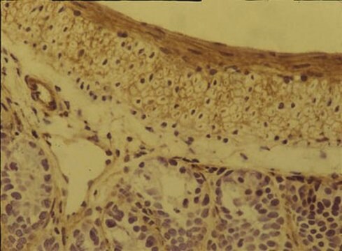
Immunohistochemistry:
1:1000 dilution of a previous lot was used in tissue staining; suggested for use on acetone fixed tissue only.

Immunocytochemistry:
1:1000 of a previous lot was used in immunocytochemistry.

Optimal working dilutions must be determined by the end user.

Quality

Routinely evaluated by Western Blot on HUVEC lysates.

Western Blot Analysis: 1:1000 dilution of this lot detected integrin alpha 5 on 10 μg of HUVEC lysates.

Target description

114 kDa

Physical form

Rabbit polyclonal antiserum in buffer containing 0.05% sodium azide.

Analysis Note

Control
Mouse 3T3 fibroblasts Skin (Basement Membrane).

Other Notes

Concentration: Please refer to the Certificate of Analysis for the lot-specific concentration.

Legal Information

CHEMICON is a registered trademark of Merck KGaA, Darmstadt, Germany

Not finding the right product?  

Try our Product Selector Tool.

Storage Class Code

12 - Non Combustible Liquids

WGK

WGK 1

Flash Point(F)

Not applicable

Flash Point(C)

Not applicable


Certificates of Analysis (COA)

Search for Certificates of Analysis (COA) by entering the products Lot/Batch Number. Lot and Batch Numbers can be found on a product’s label following the words ‘Lot’ or ‘Batch’.

Already Own This Product?

Find documentation for the products that you have recently purchased in the Document Library.

Visit the Document Library

Mary Ann Stepp et al.
Molecular carcinogenesis, 49(4), 363-373 (2010-01-19)
Syndecan-1 (sdc-1) is a cell surface proteoglycan that mediates the interaction of cells with their matrix, influencing attachment, migration, and response to growth factors. In keratinocytes, loss of sdc-1 delays wound healing, reduces migration, and increases Transforming growth factor beta
Vincent F Fiore et al.
The Journal of cell biology, 211(1), 173-190 (2015-10-16)
Progressive fibrosis is characterized by excessive deposition of extracellular matrix (ECM), resulting in gross alterations in tissue mechanics. Changes in tissue mechanics can further augment scar deposition through fibroblast mechanotransduction. In idiopathic pulmonary fibrosis, a fatal form of progressive lung
Kazuko Goto et al.
Stem cells translational medicine, 5(2), 218-226 (2015-12-25)
When injected directly into ischemic tissue in patients with peripheral artery disease, the reparative capacity of endothelial progenitor cells (EPCs) appears to be limited by their poor survival. We, therefore, attempted to improve the survival of transplanted EPCs through intravenous
Andoria Tjondrokoesoemo et al.
The Journal of biological chemistry, 291(19), 9920-9928 (2016-03-12)
Duchenne muscular dystrophy (DMD) is an X-linked recessive disease caused by mutations in the gene encoding dystrophin. Loss of dystrophin protein compromises the stability of the sarcolemma membrane surrounding each muscle cell fiber, leading to membrane ruptures and leakiness that
Jamie L Marshall et al.
Human molecular genetics, 24(7), 2011-2022 (2014-12-17)
Duchenne muscular dystrophy (DMD) is caused by mutations in the dystrophin gene that result in loss of the dystrophin-glycoprotein complex, a laminin receptor that connects the myofiber to its surrounding extracellular matrix. Utrophin, a dystrophin ortholog that is normally localized

Our team of scientists has experience in all areas of research including Life Science, Material Science, Chemical Synthesis, Chromatography, Analytical and many others.

Contact Technical Service