Skip to Content
Merck
All Photos(4)

Documents

05-175

Sigma-Aldrich

Anti-Myb Antibody, clone 1-1

clone 1-1, Upstate®, from mouse

Synonym(s):

Avian myeloblastosis viral (v-myb) oncogene homolog 2 c-myb protein (140 AA), v-myb avian myeloblastosis viral oncogene homolog, v-myb avian myeloblastosis viral oncogene homolog, v-myb myeloblastosis viral oncogene homolog, v-myb myeloblastosis viral on

Sign Into View Organizational & Contract Pricing


About This Item

UNSPSC Code:
12352203
eCl@ss:
32160702
NACRES:
NA.41

biological source

mouse

Quality Level

antibody form

purified immunoglobulin

antibody product type

primary antibodies

clone

1-1, monoclonal

species reactivity

mouse, human

manufacturer/tradename

Upstate®

technique(s)

immunohistochemistry: suitable
immunoprecipitation (IP): suitable
western blot: suitable

isotype

IgG2aκ

NCBI accession no.

UniProt accession no.

shipped in

dry ice

target post-translational modification

unmodified

Gene Information

human ... MYB(4602)

General description

Myb is a transcriptional activator. It is a DNA-binding protein that specifically recognizes the sequence 5′-YAAC[GT]G-3′. Myb plays an important role in the control of proliferation and differentiation of hematopoietic progenitor cells.

Specificity

This antibody recognizes Myb, Mr 80 kDa, containing C-terminal amino acids from 401-636 (Salomoni, P., 1997).

Immunogen

Recombinant murine Myb purified by SDS-PAGE (Nicot, C., 2001) Clone 1-1.

Application

Anti-Myb Antibody, clone 1-1 is a Mouse Monoclonal Antibody for detection of Myb also known as Avian myeloblastosis viral (v-myb) oncogene homolog 2 c-myb protein (140 AA) & has been validated in IHC, IP & WB.
Immunoprecipitation:
4 μg of previous lot immunoprecipitated Myb from Jurkat cell lysate.

Immunohistochemistry:
Research Category
Epigenetics & Nuclear Function
Research Sub Category
Transcription Factors

Quality

Routinely evaluated on RIPA lysate from Jurkat cells.

Western Blot Analysis:
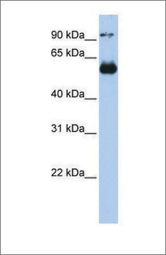
0.5-2 μg/mL of this lot detected Myb in 20 μg of RIPA lysate from Jurkat cells.

Target description

80 kDa

Physical form

Ammonium sulfate precipitation, gel filtration, and ion-exchange chromatography.
Format: Purified
Purified mouse monoclonal IgG2aκ in buffer containing 0.1 M Tris-glycine, pH 7.4, 0.15 M NaCl and 0.05% sodium azide. Frozen solution.

Storage and Stability

Stable for 1 year at -20°C from date of receipt. Aliquot to avoid repeated freezing and thawing. For maximum recovery of product, centrifuge the original vial after thawing and prior to removing the cap.

Analysis Note

Control
Positive Antigen Control: Catalog #12-303, Jurkat cell lysate.

Other Notes

Concentration: Please refer to the Certificate of Analysis for the lot-specific concentration.

Legal Information

UPSTATE is a registered trademark of Merck KGaA, Darmstadt, Germany

Disclaimer

Unless otherwise stated in our catalog or other company documentation accompanying the product(s), our products are intended for research use only and are not to be used for any other purpose, which includes but is not limited to, unauthorized commercial uses, in vitro diagnostic uses, ex vivo or in vivo therapeutic uses or any type of consumption or application to humans or animals.

Not finding the right product?  

Try our Product Selector Tool.

Storage Class Code

12 - Non Combustible Liquids

WGK

WGK 1

Flash Point(F)

Not applicable

Flash Point(C)

Not applicable


Certificates of Analysis (COA)

Search for Certificates of Analysis (COA) by entering the products Lot/Batch Number. Lot and Batch Numbers can be found on a product’s label following the words ‘Lot’ or ‘Batch’.

Already Own This Product?

Find documentation for the products that you have recently purchased in the Document Library.

Visit the Document Library

cDNA array analysis of alterations in gene expression in the promyelocytic leukemia cell line, HL-60, after apoptosis induction with etoposide.
M Bj?rling-Poulsen, O-G Issinger
Apoptosis null
c-Maf interacts with c-Myb to down-regulate Bcl-2 expression and increase apoptosis in peripheral CD4 cells.
Siying Peng,Saif Lalani,Jianmei W Leavenworth,I-Cheng Ho,Mary E Pauza
European Journal of Immunology null
Zhenglong Wu et al.
Journal of experimental & clinical cancer research : CR, 32, 98-98 (2013-12-03)
Mir-29 microRNA families are involved in regulation of various types of cancers. Although Mir-29 was shown to play an inhibitory role in tumorigenesis, the role of Mir-29 in breast cancer still remains obscure. In this study, we showed that Mir-29a
XinJing Tang et al.
Nucleic acids research, 36(2), 559-569 (2007-12-07)
Light-activated antisense oligodeoxynucleotides (asODNs) were developed to control the degradation of target mRNA in living cells by RNase H. A 20-mer asODN previously shown to target c-myb, a hematopoietic transcription factor, was covalently attached via a photocleavable linker (PL) to
Delivery of c-myb antisense oligodeoxynucleotides to human neuroblastoma cells via disialoganglioside GD(2)-targeted immunoliposomes: antitumor effects.
G Pagnan, D D Stuart, F Pastorino, L Raffaghello, P G Montaldo, T M Allen, B Calabretta, M Ponzoni
Journal of the National Cancer Institute null

Our team of scientists has experience in all areas of research including Life Science, Material Science, Chemical Synthesis, Chromatography, Analytical and many others.

Contact Technical Service