Přejít k obsahu
Merck
Všechny fotografie(3)

Key Documents

MABS1266

Sigma-Aldrich

Anti-LOX-1 Antibody, clone 15C4

clone 15C4, from mouse

Synonyma:

Oxidized low-density lipoprotein receptor 1, Ox-LDL receptor 1, C-type lectin domain family 8 member A, Lectin-like oxidized LDL receptor 1, Lectin-like oxLDL receptor 1, hLOX-1, Lectin-type oxidized LDL receptor 1

Přihlásitk zobrazení cen stanovených pro organizaci a smluvních cen


About This Item

UNSPSC Code:
12352203
eCl@ss:
32160702
NACRES:
NA.41

biological source

mouse

Quality Level

antibody form

purified immunoglobulin

antibody product type

primary antibodies

clone

15C4, monoclonal

species reactivity

human

technique(s)

flow cytometry: suitable
immunofluorescence: suitable
immunohistochemistry: suitable
western blot: suitable

isotype

IgG2aκ

NCBI accession no.

UniProt accession no.

shipped in

ambient

target post-translational modification

unmodified

Gene Information

human ... OLR1(4973)

General description

Oxidized low-density lipoprotein receptor 1 (UniProt: P78380; also known as Ox-LDL receptor 1, C-type lectin domain family 8 member A, Lectin-like oxidized LDL receptor 1, LOX-1, Lectin-like oxLDL receptor 1, hLOX-1, Lectin-type oxidized LDL receptor 1) is encoded by the OLR1 (also known as CLEC8A, LOX1) gene (Gene ID: 4973) in human. LOX1 is a type II transmembrane receptor for oxidatively modified low density lipoprotein (oxLDL), a well-known marker of atherosclerosis. A free form of LOX-1 has also been reported. LOX-1 has a The C-type lectin domain that mediates the recognition and binding of oxLDL. Its cytoplasmic region is considered to be essential for subcellular sorting on the cell surface. LOX-1 is expressed at high level in endothelial cells and vascular-rich organs such as placenta, lung, liver and brain, aortic intima, bone marrow, spinal cord and substantia nigra. It is also expressed at the surface of dendritic cells. LOX-1 mediates the recognition, internalization and degradation of oxLDL by vascular endothelial cells. OxLDL induces vascular endothelial cell activation and dysfunction, resulting in pro-inflammatory responses, pro-oxidative conditions and also apoptosis. In addition to binding oxLDL, LOX-1 also acts as a receptor for the HSP70 protein involved in antigen cross-presentation to naive T-cells in dendritic cells, thereby participating in cell-mediated antigen cross-presentation. LOX-1 is up-regulated in atherosclerotic lesions, by oxLDL, reactive oxygen species and fluid shear stress.

Specificity

Clone 15C4 recognizes LOX-1 in human cells.

Immunogen

Human LOX 1 ectodomain-hIgGFc fusion protein.

Application

Detect LOX-1 using this mouse monoclonal Anti-LOX-1 Antibody, clone 15C4, Cat. No. MABS1266. It has been tested in Cell Differentiation studies, Flow Cytometry, Immunofluorescence, Immunohistochemistry, and Western Blotting.
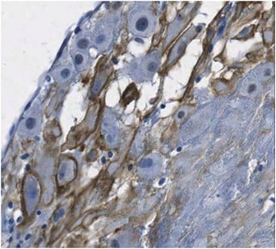
Immunohistochemistry Analysis: A 1:250-1,000 dilution from a representative lot detected LOX-1 in human spleen, human large intestine, and human vagina tissues.

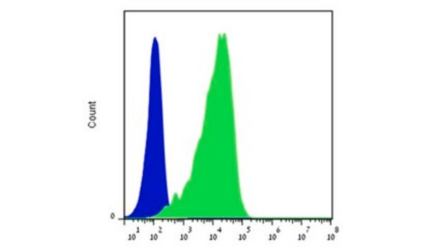
Flow Cytometry Analysis: A representative lot detected LOX-1 in Flow Cytometry applications (Li, D., et. al. (2012). J Exp Med. 209(1):109-21).

Immunofluorescence Analysis: A representative lot detected LOX-1 in Immunofluorescence applications (Li, D., et. al. (2012). J Exp Med. 209(1):109-21).

Cell Differentiation Analysis: A representative lot detected LOX-1 in Cell Differentiation applications (Joo, H., et. al. (2014). Immunity. 41(4):592-604).

Immunohistochemistry Analysis: A representative lot detected LOX-1 in Immunohistochemistry applications (Duluc, D., et. al. (2013). Microb Pathog. 58:35-44).

Flow Cytometry Analysis: 1 ug from a representative lot detected LOX-1 in one million A549 cells.
Research Category
Signaling

Quality

Evaluated by Western Blotting in human liver tissue lysates.

Western Blotting Analysis: 2 µg/mL of this antibody detected LOX-1 in 10 µg of human liver tissue lysates.

Target description

30.96 kDa calculated. Uncharacterized band(s) may appear in some lysates.

Physical form

Format: Purified
Protein G purified
Purified mouse monoclonal antibody IgG2a in PBS without preservatives.

Storage and Stability

Stable for 1 year at -20°C from date of receipt.

Handling Recommendations: Upon receipt and prior to removing the cap, centrifuge the vial and gently mix the solution. Aliquot into microcentrifuge tubes and store at -20°C. Avoid repeated freeze/thaw cycles, which may damage IgG and affect product performance.

Other Notes

Concentration: Please refer to lot specific datasheet.

Disclaimer

Unless otherwise stated in our catalog or other company documentation accompanying the product(s), our products are intended for research use only and are not to be used for any other purpose, which includes but is not limited to, unauthorized commercial uses, in vitro diagnostic uses, ex vivo or in vivo therapeutic uses or any type of consumption or application to humans or animals.

Ještě jste nenalezli správný produkt?  

Vyzkoušejte náš produkt Nástroj pro výběr produktů.

Storage Class

12 - Non Combustible Liquids

wgk_germany

WGK 2

flash_point_f

Not applicable

flash_point_c

Not applicable


Osvědčení o analýze (COA)

Vyhledejte osvědčení Osvědčení o analýze (COA) zadáním čísla šarže/dávky těchto produktů. Čísla šarže a dávky lze nalézt na štítku produktu za slovy „Lot“ nebo „Batch“.

Již tento produkt vlastníte?

Dokumenty související s produkty, které jste v minulosti zakoupili, byly za účelem usnadnění shromážděny ve vaší Knihovně dokumentů.

Navštívit knihovnu dokumentů

Náš tým vědeckých pracovníků má zkušenosti ve všech oblastech výzkumu, včetně přírodních věd, materiálových věd, chemické syntézy, chromatografie, analytiky a mnoha dalších..

Obraťte se na technický servis.