Skip to Content
Merck
All Photos(1)

Key Documents

H6412

Sigma-Aldrich

Monoclonal Anti-Histone Deacetylase 8 (HDAC8) antibody produced in mouse

2.0-2.5 mg/mL, clone HDAC8-48, purified immunoglobulin, buffered aqueous solution

Sign Into View Organizational & Contract Pricing


About This Item

MDL number:
UNSPSC Code:
12352203
NACRES:
NA.41

biological source

mouse

Quality Level

conjugate

unconjugated

antibody form

purified immunoglobulin

antibody product type

primary antibodies

clone

HDAC8-48, monoclonal

form

buffered aqueous solution

mol wt

antigen ~43 kDa

species reactivity

human

concentration

2.0-2.5 mg/mL

technique(s)

indirect ELISA: suitable
microarray: suitable
western blot: 4 μg/mL using nuclear extracts of HeLa cells

isotype

IgG1

UniProt accession no.

shipped in

dry ice

storage temp.

−20°C

target post-translational modification

unmodified

Gene Information

human ... HDAC8(55869)

General description

Histone deacetylases (HDACs) are competing enzymes, belonging to histone deacetylase family. There are two classes of HDACs with six to seven different types of HDACs proteins. HDAC1,HDAC2, HDAC3, and HDAC8 belong to Class I HDACs and HDAC4, HDAC6, HDAC7, HDAC9, and HDAC10 belong to Class II HDACs. Class I HDACs consists of a single deacetylase domain at the N-termini and diversified C-terminal regions, while Class II contains a deacetylase domain at C-terminal position. Studies show that HDAC8 is a sex-linked gene located on the chromosome at position Xq21.2 - q21.3. HDAC8 gene has a molecular weight of 43kDa and it encodes a 377 amino acid protein. It is present within the nucleas. HDAC8 mRNA is seen in heart, lung, kidney, and pancreas.
Monoclonal Anti-Histone Deacetylase 8 (HDAC8) (mouse IgG1 isotype) is derived from the HDAC8-48 hybridoma produced by the fusion of NS-1 mouse myeloma cells and splenocytes from BALB/c mice immunized with recombinant human HDAC8.

Immunogen

recombinant human HDAC8

Application

Monoclonal Anti-Histone Deacetylase 8 (HDAC8) antibody produced in mouse has been used in :
  • enzyme linked immunosorbent assay (ELISA)
  • immunoblotting
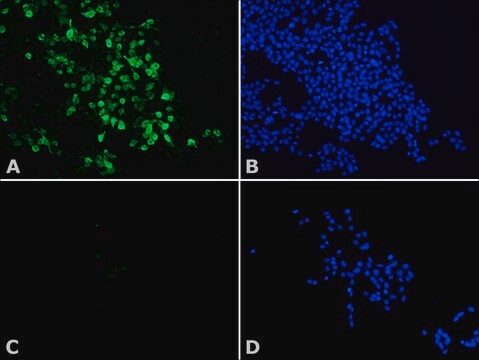
  • immunofluorescence staining

Biochem/physiol Actions

Histone deacetylation results in transcription repression leading to the formation of tight nucleosomal structure which prevents DNA accessing. HDAC8 controls HDAC activity on H4 histone peptide substrates. HDAC8 is similar to the HDAC class I enzymes. In vitro expression and activity of HDAC8 was examined using FLAG tagged- HDAC8 and HDAC1. These were transfected into HeLa cells and Sf9 insect cells. After the immunoprecipitation of cell lysates the expression results were confirmend using Western blotting. Studies show that HDAC8 may play a role in transcriptional regulation and could possibly be regulated in a temporal or compartment-specific manner.

Physical form

Solution in 0.01 M phosphate buffered saline, pH 7.4, containing 15 mM sodium azide.

Disclaimer

Unless otherwise stated in our catalog or other company documentation accompanying the product(s), our products are intended for research use only and are not to be used for any other purpose, which includes but is not limited to, unauthorized commercial uses, in vitro diagnostic uses, ex vivo or in vivo therapeutic uses or any type of consumption or application to humans or animals.

Not finding the right product?  

Try our Product Selector Tool.


Certificates of Analysis (COA)

Search for Certificates of Analysis (COA) by entering the products Lot/Batch Number. Lot and Batch Numbers can be found on a product’s label following the words ‘Lot’ or ‘Batch’.

Already Own This Product?

Find documentation for the products that you have recently purchased in the Document Library.

Visit the Document Library

An atlas of histone deacetylase expression in breast cancer: fluorescence methodology for comparative semi-quantitative analysis
Ververis K and Karagiannis TC
American Journal of Translational Research, 4(1), 24-24 (2012)
Satoshi Inoue et al.
Cancer research, 66(13), 6785-6792 (2006-07-05)
From work done largely on derived cell lines, it has been suggested that tumor necrosis factor-related apoptosis-inducing ligand (TRAIL) might be a therapeutic target for many forms of malignancy. However, use of primary tumor cells, including chronic lymphocytic leukemic (CLL)
Inhibition of histone deacetylases 1 and 6 enhances cytarabine-induced apoptosis in pediatric acute myeloid leukemia cells
Xu X, et al.
Testing, 6(2), e17138-e17138 (2011)
Katherine Ververis et al.
American journal of translational research, 4(1), 24-43 (2012-02-22)
The histone deacetylase inhibitors, suberoylanilide hydroxamic acid (Vorinostat, Zolinza™) and depsipeptide (Romidepsin, Istodax™) have been approved by the US Food and Drug Administration for the treatment of refractory cutaneous T-cell lymphoma. Numerous histone deacetylase inhibitors are currently undergoing clinical trials
Xuelian Xu et al.
PloS one, 6(2), e17138-e17138 (2011-03-02)
Pediatric acute myeloid leukemia (AML) remains a challenging disease to treat even with intensified cytarabine-based chemotherapy. Histone deacetylases (HDACs) have been reported to be promising therapeutic targets for treating AML. However, HDAC family members that are involved in chemotherapy sensitivities

Our team of scientists has experience in all areas of research including Life Science, Material Science, Chemical Synthesis, Chromatography, Analytical and many others.

Contact Technical Service