Skip to Content
Merck
All Photos(1)

Key Documents

MAB1562

Sigma-Aldrich

Anti-Prion Protein Antibody, a.a. 109-112, clone 3F4

clone 3F4, Chemicon®, from mouse

Synonym(s):

PrP, CD230

Sign Into View Organizational & Contract Pricing


About This Item

UNSPSC Code:
12352203
eCl@ss:
32160702
NACRES:
NA.41

biological source

mouse

Quality Level

antibody form

purified immunoglobulin

antibody product type

primary antibodies

clone

3F4, monoclonal

species reactivity

hamster, human

manufacturer/tradename

Chemicon®

technique(s)

ELISA: suitable
immunohistochemistry (formalin-fixed, paraffin-embedded sections): suitable
immunoprecipitation (IP): suitable
western blot: suitable

isotype

IgG2a

NCBI accession no.

UniProt accession no.

shipped in

dry ice

target post-translational modification

unmodified

Gene Information

human ... PRNP(5621)

General description

Prions are thought to cause a number of diseases in a variety of mammals, including bovine spongiform encephalopathy (BSE, also known as "mad cow disease") in cattle and the Creutzfeldt-Jakob disease (CJD) in humans. All thus-far hypothesized prion diseases affect the structure of the brain or other neural tissue, and all are currently untreatable and thought to be fatal. Prions are hypothesized to infect and propagate by refolding abnormally into a structure which is able to convert normal molecules of the protein into the abnormally structured form. All known prions induce the formation of an amyloid fold, in which the protein polymerises into an aggregate consisting of tightly packed beta sheets. This altered structure is extremely stable and accumulates in infected tissue, causing cell death and tissue damage. This stability means that prions are resistant to denaturation by chemical and physical agents, making disposal and containment of these particles difficult.

Specificity

Prion protein, amino acid residues 109-112 of human, hamster and feline. Does not react with PrP from any other mammalian species. MAB1562 is reactive to native and denatured forms of PrP. Tissue or cells which have been fixed requires that the epitope be re-exposed (see below). Recognizes both protease sensitive and protease resistant forms of PrP.

Immunogen

Epitope: a.a. 109-112

Application

Immunohistochemistry(paraffin):
Representative images from a previous lot. Optimal Staining With Citrate Buffer, pH 6.0, Epitope Retrieval: Human Brain

Immunohistochemistry (Kitamoto et al., 1987):
1:100-1:1,000 *See protocol below.

Epitope must be re-exposed in fixed tissue by pretreatment of tissue using one of the following procedures:
a. formic acid for 10 minutes at room temperature (Kitamoto et al., 1987)
b. hydrolytic autoclaving (Kitamoto et al., 1991)
c. microwaving (BioGenex, San Ramon, CA)

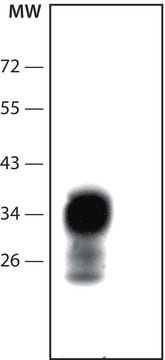
Western Blot: (Kascsak, R.J., 1991; Kascsak, R.J., 1987):
1:10,000-1:100,000 dilution of a previous lot was used.

Immunoprecipitation: (Kascsak, R.J., 1991; Kascsak, R.J., 1987):
1:10-1:100 dilution of a previous lot was used.

ELISA: (Kascsak, R.J., 1991; Kascsak, R.J., 1987):
1:100,000 dilution of a previous lot was used.

Optimal working dilutions must be determined by end user.
This Anti-Prion Protein Antibody, a.a. 109-112, clone 3F4 is validated for use in ELISA, IH, IH(P), IP, WB for the detection of Prion Protein.

Quality

Immunohistochemistry(paraffin):
Prion Protein (cat. # MAB1562) staining pattern/morphology in normal brain. Tissue was pretreated with Citrate, pH 6.0. This lot of antibody was diluted to 1:500, using IHC-Select Detection with HRP-DAB. Immunoreactivity is seen predominantly as cell body staining of neurons.
Optimal Staining With Citrate Buffer, pH 6.0, Epitope Retrieval: Human Brain

Target description

12.3 kDa

Physical form

Format: Purified
Purified mouse monoclonal IgG2a in buffer containing PBS and no preservative.

Legal Information

CHEMICON is a registered trademark of Merck KGaA, Darmstadt, Germany

Not finding the right product?  

Try our Product Selector Tool.

Storage Class Code

12 - Non Combustible Liquids

WGK

WGK 2

Flash Point(F)

Not applicable

Flash Point(C)

Not applicable


Certificates of Analysis (COA)

Search for Certificates of Analysis (COA) by entering the products Lot/Batch Number. Lot and Batch Numbers can be found on a product’s label following the words ‘Lot’ or ‘Batch’.

Already Own This Product?

Find documentation for the products that you have recently purchased in the Document Library.

Visit the Document Library

Qi Shi et al.
International journal of molecular medicine, 41(4), 2413-2419 (2018-02-03)
Normal prion protein (PrP) contains two cysteines at amino acids 179 and 214, which may form intra‑ and interpeptide disulfide bonds. To determine the possible effects of this disulfide bridge on the biochemical features of PrP, prokaryotic recombinant human wild‑type PrP
Michele Christine Landemberger et al.
Journal of neurochemistry, 145(5), 409-416 (2018-01-18)
Cellular prion protein (PrPC ) is widely expressed and displays a variety of well-described functions in the central nervous system (CNS). Mutations of the PRNP gene are known to promote genetic human spongiform encephalopathies, but the components of gain- or
Zuzana Krejciova et al.
The Journal of experimental medicine, 214(12), 3481-3495 (2017-11-17)
Prions are infectious agents that cause neurodegenerative diseases such as Creutzfeldt-Jakob disease (CJD). The absence of a human cell culture model that replicates human prions has hampered prion disease research for decades. In this paper, we show that astrocytes derived
Pol Andres Benito et al.
Prion, 12(3-4), 216-225 (2018-07-17)
Targeted expression of genes coding for proteins specific to astrocytes, oligodendrocytes and myelin was performed in frontal cortex area 8 of Creutzfeldt-Jakob disease methionine/methionine and valine/valine (CJD MM1 and VV2, respectively) compared with controls. GFAP (glial fibrillary acidic protein) mRNA
Francesca Properzi et al.
The Journal of general virology, 96(Pt 7), 1969-1974 (2015-03-26)
In most forms of prion diseases, blood is infectious, but detection by immunochemistry techniques of the only available marker of infection (the misfolded prion protein, PrPTSE) in blood remains elusive. We developed a novel method for the detection of PrPTSE

Our team of scientists has experience in all areas of research including Life Science, Material Science, Chemical Synthesis, Chromatography, Analytical and many others.

Contact Technical Service