Recommended Products
biological source
rabbit
Quality Level
antibody form
affinity isolated antibody
antibody product type
primary antibodies
clone
polyclonal
form
liquid
does not contain
preservative
species reactivity
mouse, rat, human
manufacturer/tradename
Calbiochem®
storage condition
OK to freeze
avoid repeated freeze/thaw cycles
isotype
IgG
shipped in
wet ice
storage temp.
−20°C
target post-translational modification
unmodified
Gene Information
human ... MAPK8(5599)
General description
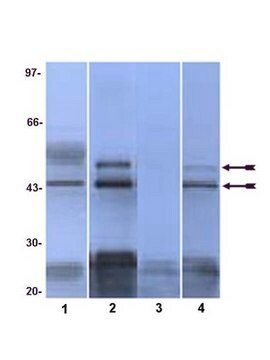
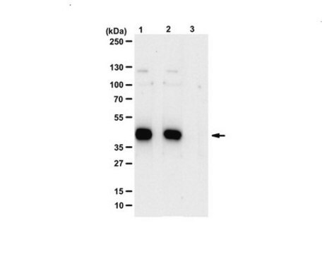
Protein A and immunoaffinity purified rabbit polyclonal antibody. Recognizes the ~54 kDa SAPK/JNK protein.
Recognizes the ~54 kDa SAPK/JNK protein in uv-treated HEK293 cells.
This Anti-SAPK/JNK Rabbit pAb is validated for use in Immunoblotting, Immunocytochemistry for the detection of SAPK/JNK.
Immunogen
Human
a full-length, recombinant, human p54 SAPK/JNK2 fusion protein
Application
Immunoblotting (1:1000)
Immunocytochemistry (1:200)
Immunocytochemistry (1:200)
Warning
Toxicity: Standard Handling (A)
Physical form
In 150 mM NaCl, 10 mM HEPES, 50% glycerol, 0.01% BSA, pH 7.5.
Reconstitution
Following initial thaw, aliquot and freeze (-20°C).
Analysis Note
Positive Control
UV treated HEK293 cells
UV treated HEK293 cells
Other Notes
Gupta, S., et al. 1996. EMBO J.15(11), 2760.
Coso, O.A., et al. 1995. Cell81, 1137.
Derijard, B., et al. 1994. Cell76, 1025.
Kyriakis, J.M., et al. 1994. Nature369, 156.
Hibi, M., et al. 1993. Genes Dev.7, 2135.
Kyriakis, J.M. and Avruch, J. 1990. J. Biol. Chem.265, 17355.
Coso, O.A., et al. 1995. Cell81, 1137.
Derijard, B., et al. 1994. Cell76, 1025.
Kyriakis, J.M., et al. 1994. Nature369, 156.
Hibi, M., et al. 1993. Genes Dev.7, 2135.
Kyriakis, J.M. and Avruch, J. 1990. J. Biol. Chem.265, 17355.
Recognizes SAPK/JNK regardless of the phosphorylation state. Variables associated with assay conditions will dictate the proper working dilution.
Recommended Protocol for Immunoblotting
Solutions and Reagents
•Transfer Buffer: 25 mM Tris base, 0.2 M glycine, 20% methanol, pH 8.5.
•SDS Sample Buffer: 62.5 mM Tris-HCl, pH 6.8, 2% SDS, 10% glycerol, 50 mM DTT, 0.1% bromphenol blue.
•10X TBS (Tris-buffered saline): To prepare 1 liter, 24.2 g Tris base, 80 g NaCl, adjust pH to 7.6 with HCl. Dilute 1:10 for use.
•Blocking Buffer: 1X TBS, 0.1% Tween®-20 detergent with 5% non-fat dry milk.
•Primary Antibody Dilution Buffer: 1X TBS, 0.1% Tween-20 detergent with 5% BSA
•Wash Buffer (TBST): 1X TBS, 0.1% Tween-20 detergent
Blotting Membrane
Nitrocellulose or PVDF membranes may be used.
Protein Blotting
A general protocol for sample preparation using 2x10⁶ 293 cells per well in a 6-well plate is as follows:
1. Aspirate media. Treat cells by adding fresh media containing regulator for desired time.
2. Aspirate media from cultures; wash cells with PBS; aspirate.
3. Lyse cells by adding 100 µl of SDS Sample Buffer and immediately scrape the cells off the plate and transfer the extract to a microfuge tube. Keep on ice.
4. Sonicate for 2 s to shear DNA and reduce sample viscosity.
5. Heat sample to 95-100°C for 5 min. Cool on ice.
6. Microcentrifuge for 5 min.
7. Load 20 µl onto SDS-PAGE gel (10 cm x 10 cm).
8. Electrotransfer to nitrocellulose membrane.
As controls, we recommend using 20 µl lysate from UV treated HEK293 cell.
Membrane Blocking, Gel and Antibody Incubations
1. After transfer, wash membrane with 25 ml TBS for 5 min at room temperature.
2. Incubate membrane in 25 ml Blocking Buffer for 1-3 h at room temperature or overnight at 4°C.
3. Wash 3 times for 5 min each with 15 ml TBST.
4. Incubate membrane and primary antibody (at the appropriate dilution) in 10 ml Primary Antibody Dilution Buffer with gentle agitation overnight at 4°C.
5. Wash 3 times for 5 min each with 15 ml TBST.
6. Incubate membrane with conjugated secondary antibody at the appropriate dilution in 10 ml Blocking Buffer with gentle agitation for 1 h at room temperature.
7. Wash membrane as in step 5.
Detection of Proteins
Chemiluminescence.
Legal Information
CALBIOCHEM is a registered trademark of Merck KGaA, Darmstadt, Germany
TWEEN is a registered trademark of Croda International PLC
Not finding the right product?
Try our Product Selector Tool.
Storage Class Code
10 - Combustible liquids
WGK
WGK 1
Certificates of Analysis (COA)
Search for Certificates of Analysis (COA) by entering the products Lot/Batch Number. Lot and Batch Numbers can be found on a product’s label following the words ‘Lot’ or ‘Batch’.
Already Own This Product?
Find documentation for the products that you have recently purchased in the Document Library.
Peter Verstraelen et al.
Cellular and molecular gastroenterology and hepatology, 18(1), 89-104 (2024-04-01)
Mounting evidence suggests the gastrointestinal microbiome is a determinant of peripheral immunity and central neurodegeneration, but the local disease mechanisms remain unknown. Given its potential relevance for early diagnosis and therapeutic intervention, we set out to map the pathogenic changes
Vincenzo Giansanti et al.
Journal of cellular and molecular medicine, 17(1), 103-115 (2012-12-05)
The pathogenesis of age-related macular degeneration (AMD) involves demise of the retinal pigment epithelium and death of photoreceptors. In this article, we investigated the response of human adult retinal pigmented epithelial (ARPE-19) cells to 5-(N,N-hexamethylene)amiloride (HMA), an inhibitor of Na(+)
Kyubin Lee et al.
Biology, 9(8) (2020-08-13)
Abeliophyllum distichum Nakai is known as a monotypic genus endemic to South Korea. Currently, several pharmacological studies have revealed that A. distichum extract exhibits diverse biological functions, including anti-cancer, anti-diabetic, anti-hypertensive, and anti-inflammatory activities. In this study, we present the
Aveline Filliol et al.
Scientific reports, 7(1), 9205-9205 (2017-08-25)
Hepatocyte death is a central event during liver disease progression, in which immune cells play key roles by activating members of the Tumor Necrosis Factor Receptor Superfamily (TNFRSF), including TNFR1 (TNFRSF1A), Fas (TNFRSF6) and TRAIL-R2 (TNFRSF10B). Receptor Interacting Protein Kinase
Our team of scientists has experience in all areas of research including Life Science, Material Science, Chemical Synthesis, Chromatography, Analytical and many others.
Contact Technical Service


