Saltar al contenido
Merck
Todas las fotos(9)

Key Documents

HPA010800

Sigma-Aldrich

Anti-DCT antibody produced in rabbit

enhanced validation

Prestige Antibodies® Powered by Atlas Antibodies, affinity isolated antibody, buffered aqueous glycerol solution

Sinónimos:

Anti-DCT, Anti-DT, Anti-L- dopachrome Delta-isomerase, Anti-L-dopachrome tautomerase precursor, Anti-TRP-2, Anti-TRP2, Anti-Tyrosinase-related protein 2

Iniciar sesiónpara Ver la Fijación de precios por contrato y de la organización


About This Item

UNSPSC Code:
12352203
Human Protein Atlas Number:
NACRES:
NA.43

biological source

rabbit

Quality Level

conjugate

unconjugated

antibody form

affinity isolated antibody

antibody product type

primary antibodies

clone

polyclonal

product line

Prestige Antibodies® Powered by Atlas Antibodies

form

buffered aqueous glycerol solution

species reactivity

human

enhanced validation

independent
orthogonal RNAseq
recombinant expression
Learn more about Antibody Enhanced Validation

technique(s)

immunoblotting: 0.04-0.4 μg/mL
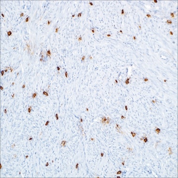
immunohistochemistry: 1:200-1:500

immunogen sequence

NECDVCTDQLFGAARPDDPTLISRNSRFSSWETVCDSLDDYNHLVTLCNGTYEGLLRRNQMGRNSMKLPTLKDIRDCLSLQKFDNPPFFQNSTFSFRNALEGFDKADGTLDSQVMSLHNLVHSFLNGTNALPHSAANDPIFVVLHSFTDA

UniProt accession no.

shipped in

wet ice

storage temp.

−20°C

target post-translational modification

unmodified

Gene Information

human ... DCT(1638)

General description

DCT (dopachrome tautomerase) belongs to melanocyte/melanoma differentiation antigens (MDA) family. It is a melanoma associated antigen. It contains 519 amino acids and has five isoforms in humans, due to alternative splicing. It contains a putative transmembrane domain, encoded by exon 8, which is essential for its activity. TRP (tyrosinase-related protein)-LT and TRP-2-6b isoforms of DCT are localized to the membrane of melanosomes, and possess dopachrome tautomerase activity. TRP-2-INT2 isoform is a shorter protein with only 237 amino acids. TRP- 2-8b isoform does not contain the transmembrane domain, and hence is devoid of dopachrome tautomerase activity. This gene is located on human chromosome 13q32.

Immunogen

L-dopachrome tautomerase precursor recombinant protein epitope signature tag (PrEST)

Application

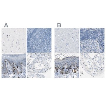
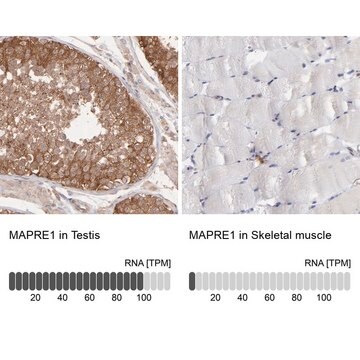
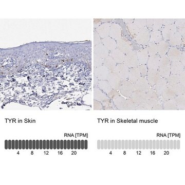
All Prestige Antibodies Powered by Atlas Antibodies are developed and validated by the Human Protein Atlas (HPA) project and as a result, are supported by the most extensive characterization in the industry.

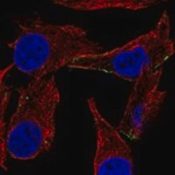
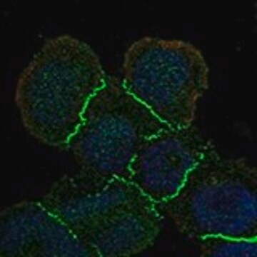
The Human Protein Atlas project can be subdivided into three efforts: Human Tissue Atlas, Cancer Atlas, and Human Cell Atlas. The antibodies that have been generated in support of the Tissue and Cancer Atlas projects have been tested by immunohistochemistry against hundreds of normal and disease tissues and through the recent efforts of the Human Cell Atlas project, many have been characterized by immunofluorescence to map the human proteome not only at the tissue level but now at the subcellular level. These images and the collection of this vast data set can be viewed on the Human Protein Atlas (HPA) site by clicking on the Image Gallery link. We also provide Prestige Antibodies® protocols and other useful information.

Biochem/physiol Actions

DCT (dopachrome tautomerase) acts as an antigen to cytotoxic T-cell responses, in melanoma. It also acts as an antigen to cytotoxic T-cells in malignant glioma. Thus, it has potential as a target antigen for developing immunotherapies for glioma patients. Polymorphisms in this gene are linked to increased susceptibility to degenerative disc disease, in Chinese Han population. DCT is absent in the peripheral blood mononuclear cells (PBMC) of vitiligo patients, but is present in low levels in healthy individuals. This lack of DCT in PBMC of vitiligo patients might be responsible for peripheral intolerance in vitiligo. It also prevents dopamine and hydroquinone-induced toxicity in human embryonic kidney (HEK) cells.

Features and Benefits

Prestige Antibodies® are highly characterized and extensively validated antibodies with the added benefit of all available characterization data for each target being accessible via the Human Protein Atlas portal linked just below the product name at the top of this page. The uniqueness and low cross-reactivity of the Prestige Antibodies® to other proteins are due to a thorough selection of antigen regions, affinity purification, and stringent selection. Prestige antigen controls are available for every corresponding Prestige Antibody and can be found in the linkage section.

Every Prestige Antibody is tested in the following ways:
  • IHC tissue array of 44 normal human tissues and 20 of the most common cancer type tissues.
  • Protein array of 364 human recombinant protein fragments.

Linkage

Corresponding Antigen APREST71670

Physical form

Solution in phosphate-buffered saline, pH 7.2, containing 40% glycerol and 0.02% sodium azide

Legal Information

Prestige Antibodies is a registered trademark of Merck KGaA, Darmstadt, Germany

Disclaimer

Unless otherwise stated in our catalog or other company documentation accompanying the product(s), our products are intended for research use only and are not to be used for any other purpose, which includes but is not limited to, unauthorized commercial uses, in vitro diagnostic uses, ex vivo or in vivo therapeutic uses or any type of consumption or application to humans or animals.

¿No encuentra el producto adecuado?  

Pruebe nuestro Herramienta de selección de productos.

Storage Class

10 - Combustible liquids

wgk_germany

WGK 1

flash_point_f

Not applicable

flash_point_c

Not applicable

ppe

Eyeshields, Gloves, multi-purpose combination respirator cartridge (US)


Certificados de análisis (COA)

Busque Certificados de análisis (COA) introduciendo el número de lote del producto. Los números de lote se encuentran en la etiqueta del producto después de las palabras «Lot» o «Batch»

¿Ya tiene este producto?

Encuentre la documentación para los productos que ha comprado recientemente en la Biblioteca de documentos.

Visite la Librería de documentos

Richard A Sturm et al.
Pigment cell & melanoma research, 22(5), 544-562 (2009-07-22)
The presence of melanin pigment within the iris is responsible for the visual impression of human eye colouration with complex patterns also evident in this tissue, including Fuchs' crypts, nevi, Wolfflin nodules and contraction furrows. The genetic basis underlying the
M Bam et al.
Scandinavian journal of immunology, 69(4), 366-373 (2009-03-17)
Tolerance is achieved by mechanisms occurring in both the thymus and periphery. Several reports have shown that presence of an antigen in the peripheral circulation results in tolerance induction. These reports imply that absence of a self-antigen can lead to
Hai-feng Song et al.
Zhonghua yi xue za zhi, 90(3), 148-152 (2010-04-02)
To investigate the association between Trp2 allele polymorphism with degenerative disc disease (DDD) in a Chinese Han population. A total of 125 DDD patients (58 males and 67 females, 51.8 +/- 7.6 years old), and 125 controls matched in sex
Q Michard et al.
Free radical biology & medicine, 45(7), 1002-1010 (2008-08-05)
We previously reported that melanogenic enzyme TRP-2 (or DCT for DOPAchrome tautomerase) expression in WM35 melanoma cells resulted in increased intracellular GSH levels, reduction in DNA damage induced by free radicals, and decreased cell sensitivity to oxidative stress. These effects
Gentao Liu et al.
Journal of immunotherapy (Hagerstown, Md. : 1997), 26(4), 301-312 (2003-07-05)
Tyrosinase-related protein (TRP)-2 is an immunogenic antigen in melanoma. The authors sought to investigate whether TRP-2 could be a potential target for patients with malignant glioma. RT-PCR analysis demonstrated that TRP-2 was present in 51.2% of primary tumor cell lines

Nuestro equipo de científicos tiene experiencia en todas las áreas de investigación: Ciencias de la vida, Ciencia de los materiales, Síntesis química, Cromatografía, Analítica y muchas otras.

Póngase en contacto con el Servicio técnico