Saltar al contenido
Merck
Todas las fotos(2)

Documentos clave

MABC536

Sigma-Aldrich

Anti-Vasohibin-2, clone 5E3 Antibody

clone 5E3, 1 mg/mL, from mouse

Sinónimos:

Vasohibin-2, Vasohibin-like protein

Iniciar sesiónpara Ver la Fijación de precios por contrato y de la organización


About This Item

UNSPSC Code:
12352203
eCl@ss:
32160702
NACRES:
NA.41

biological source

mouse

Quality Level

antibody form

purified immunoglobulin

antibody product type

primary antibodies

clone

5E3, monoclonal

species reactivity

human, mouse

concentration

1 mg/mL

technique(s)

immunohistochemistry: suitable
western blot: suitable

isotype

IgG1κ

NCBI accession no.

UniProt accession no.

shipped in

wet ice

target post-translational modification

unmodified

Gene Information

human ... VASH2(79805)

General description

Vasohibin-2, also known as Vasohibin-like protein, and encoded by the gene VASH2/VASHL, is a developmental regulator protein of angiogenesis induced by VEGF and present in various embryonic organs at six to 12 twelve weeks of development in humans. Vasohibin-2 acts as an angiogenesis inhibitor and is thought to help regulate organ vessel formation and maturation. In cancer, Vascohibin-2 appears to act to promote angiogenesis and is expressed in many cancers. In cancer cells, there are two isoforms. VASH2 (with 355 amino acid residues) is located in the cytoplasm while VASH2 (with 311 amino acid residues) is located in the nucleus. The former was found to be a relatively low abundance protein.

Immunogen

KLH-conjugated linear peptide corresponding to human Vasohibin 2.

Application

Anti-Vasohibin-2 antibody, clone 5E3 is an antibody against Vasohibin-2 for use in western blotting & IHC.
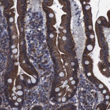
Immunohistochemistry Analysis: A 1:2,000 dilution from a representative lot detected Vasohibin 2 in human placenta tissue.
Immunohistochemistry Analysis: A representative lot detected Vasohibin 2 in human and mouse embryonic lung tissue (Shibuya, T., et al. (2006). American Heart Association. 26:1051-1057).

Quality

Evaluated by Western Blotting in HUVEC cell lysate.

Western Blotting Analysis: 0.5 µg/mL of this antibody detected Vasohibin 2 in 10 µg of HUVEC cell lysate.

Target description

~40 kDa observed. Uncharacterized band(s) may appear in some lysates.

Physical form

Format: Purified
Purified mouse monoclonal IgG1κ in buffer containing PBS without preservatives .

¿No encuentra el producto adecuado?  

Pruebe nuestro Herramienta de selección de productos.

Storage Class

12 - Non Combustible Liquids

wgk_germany

WGK 2

flash_point_f

Not applicable

flash_point_c

Not applicable


Certificados de análisis (COA)

Busque Certificados de análisis (COA) introduciendo el número de lote del producto. Los números de lote se encuentran en la etiqueta del producto después de las palabras «Lot» o «Batch»

¿Ya tiene este producto?

Encuentre la documentación para los productos que ha comprado recientemente en la Biblioteca de documentos.

Visite la Librería de documentos

Trevor J Mathias et al.
Cancers, 14(7) (2022-04-13)
Post-translational modifications (PTMs) of the microtubule network impart differential functions across normal cell types and their cancerous counterparts. The removal of the C-terminal tyrosine of α-tubulin (deTyr-Tub) as performed by the tubulin carboxypeptidase (TCP) is of particular interest in breast
Siem van der Laan et al.
Cell reports, 29(12), 4159-4171 (2019-12-19)
The two related members of the vasohibin family, VASH1 and VASH2, encode human tubulin detyrosinases. Here we demonstrate that, in contrast to VASH1, which requires binding of small vasohibin binding protein (SVBP), VASH2 has autonomous tubulin detyrosinating activity. Moreover, we

Nuestro equipo de científicos tiene experiencia en todas las áreas de investigación: Ciencias de la vida, Ciencia de los materiales, Síntesis química, Cromatografía, Analítica y muchas otras.

Póngase en contacto con el Servicio técnico