Skip to Content
Merck
All Photos(2)

Documents

SAB4200784

Sigma-Aldrich

Anti-Cellular Fibronectin antibody, Mouse monoclonal

clone FN-3E2, hybridoma cell culture supernatant

Synonym(s):

Anti-CIG, Anti-Cold-insoluble globulin, Anti-FN

Sign Into View Organizational & Contract Pricing


About This Item

UNSPSC Code:
12352203
NACRES:
NA.41

biological source

mouse

Quality Level

antibody form

culture supernatant

antibody product type

primary antibodies

clone

FN-3E2, monoclonal

form

buffered aqueous solution

mol wt

~220-260 kDa

species reactivity

chicken, rat, human, mouse

technique(s)

immunoblotting: suitable
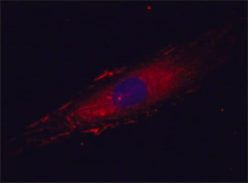
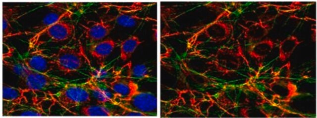
immunofluorescence: 1:2,000-1:4,000 using human foreskin fibroblast Hs68 cells
immunohistochemistry: suitable

isotype

IgM

UniProt accession no.

shipped in

dry ice

storage temp.

−20°C

target post-translational modification

unmodified

Gene Information

human ... FN1(2335)

General description

Anti-Cellular Fibronectin antibody, Mouse monoclonal (mouse IgM isotype) is derived from the FN-3E2 hybridoma produced by the fusion of mouse myeloma cells and splenocytes from mice immunized with the antigens released in culture from a breast cancer cell line. Fibronectin (FN), also known as cold-insoluble globulin (CIG), is an extracellular matrix multi-domain glycoprotein composed of two nearly identical disulphide bound polypeptides. Fibronectin 1 is a glycoprotein, that is coded by FN1 gene. It is expressed in the plasma and at the cell surface. It is mapped to human chromosome 2q35. Cellular fibronectin is produced by various cell types, including fibroblasts, endothelial cells, chondrocytes, synovial cells, and myocytes.

Immunogen

Antigens released in culture from a breast cancer cell line

Application

Anti-Cellular Fibronectin antibody, Mouse monoclonal has been used in:
  • immunoblotting
  • immunofluorescence
  • immunohistochemistry

Anti-cellular Fibronectin antibody, mouse monocloneal has been used in various immunochemical techniques including immunoblotting (220-260 kDa), immunofluorescence, and immunohistochemistry.

Biochem/physiol Actions

Fibronectin participates in cell adhesion, growth, migration, wound healing, blood coagulation and metastasis. Mutations in FN1 results in glomerulopathy. It plays an important role in cell attachment and spreading, control of cell cytoskeleton, morphology and differentiation. FN1 is also involved in extracellular matrix formation, haemostasis and thrombosis. It is a ubiquitous and essential component of the extracellular matrix (ECM) and plays a vital role in tissue repair mechanism.

Physical form

The product is supplied as a culture supernatant solution containing 15 mM sodium azide as a preservative. The product contains fetal calf serum.

Other Notes

This product is for R&D use only, not for drug, household, or other uses.

Not finding the right product?  

Try our Product Selector Tool.

Storage Class Code

10 - Combustible liquids

WGK

WGK 3

Flash Point(F)

Not applicable

Flash Point(C)

Not applicable


Certificates of Analysis (COA)

Search for Certificates of Analysis (COA) by entering the products Lot/Batch Number. Lot and Batch Numbers can be found on a product’s label following the words ‘Lot’ or ‘Batch’.

Already Own This Product?

Find documentation for the products that you have recently purchased in the Document Library.

Visit the Document Library

Plasma and cellular fibronectin: distinct and independent functions during tissue repair
To WS, et al.
Fibrogenesis & Tissue Repair, 4(1), 21-21 (2011)
The extracellular matrix proteins laminin and fibronectin contain binding domains for human plasminogen and tissue plasminogen activator.
Moser TL, et al.
The Journal of Biological Chemistry, 268(25), 18917-18923 (1993)
Receptors for cold-insoluble globulin (plasma fibronectin) on human monocytes.
Bevilacqua MP, et al.
The Journal of Experimental Medicine, 153(1), 42-60 (1981)
Paulina Escandon et al.
International journal of molecular sciences, 23(8) (2022-04-24)
Salivary exosomes have demonstrated vast therapeutic and diagnostic potential in numerous diseases. This study pioneers previously unexplored roles of SE in the context of corneal wound healing by utilizing primary corneal stromal cells from healthy (HCFs), type I diabetes mellitus
Somshuvra Bhattacharya et al.
Frontiers in bioengineering and biotechnology, 8, 1040-1040 (2020-10-06)
Oxygen deprivation within tumors is one of the most prevalent causes of resilient cancer cell survival and increased immune evasion in breast cancer (BCa). Current in vitro models do not adequately mimic physiological oxygen levels relevant to breast tissue and

Our team of scientists has experience in all areas of research including Life Science, Material Science, Chemical Synthesis, Chromatography, Analytical and many others.

Contact Technical Service