Skip to Content
Merck
All Photos(2)

Key Documents

C8736

Sigma-Aldrich

Anti-Cofilin antibody produced in rabbit

IgG fraction of antiserum, buffered aqueous solution

Synonym(s):

Anti-CFL, Anti-HEL-S-15, Anti-cofilin

Sign Into View Organizational & Contract Pricing


About This Item

MDL number:
UNSPSC Code:
12352203
NACRES:
NA.46

biological source

rabbit

Quality Level

conjugate

unconjugated

antibody form

IgG fraction of antiserum

antibody product type

primary antibodies

clone

polyclonal

form

buffered aqueous solution

mol wt

antigen ~19 kDa

species reactivity

canine, rat, human, mouse

technique(s)

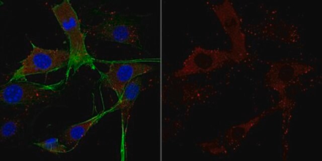
indirect immunofluorescence: 1:1,000 using using mouse NIH/3T3 fibroblasts
microarray: suitable
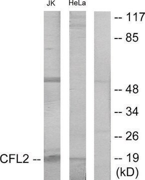
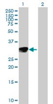
western blot: 1:10,000 using using whole extracts of human A-431, rat PC-12, mouse NIH/3T3, and dog MDCK kidney cells

UniProt accession no.

shipped in

dry ice

storage temp.

−20°C

target post-translational modification

unmodified

Gene Information

General description

Cofilin is a member of the actin depolymerizing factor/cofilin family. Among all members, cofilin-1 is the predominant isoform. It is located on human chromosome 11q13.
Cofilin is a small phosphoinositide-sensitive actin-binding protein capable of depolymerizing actin-filaments in vitro. In mammals it has two isoforms: non-muscle (NM-CF, CF-L1) and muscle (M-CF, CF-L2). The protein is ubiquitiously present in tissues of eukaryotes and is especially abundant in neuronal tissues. It can shuttle between the cytoplasm and the nucleus in response to various stresses or signals, and may translocate from the cytoplasm to the plasma membrane in various cells.

Specificity

Reacts with muscle and non-muscle cofilin.

Immunogen

Synthetic peptide corresponding to human cofilin sequence, with N-terminal cysteine added, conjugated to KLH. The corresponding sequence is identical in pig and rat non-muscle cofilin and differs by three amino acids from that of human and chicken muscle cofilin.

Application

Anti-Cofilin antibody has been used:
  • for immunostaining of chick neurons. It is used as a primary antibody
  • for western blotting of protein isolated from mouse hippocampi cells, rat brain samples, human acute lymphoblastic T-cell line, human brain samples, head and neck squamous cell carcinoma cell line, HEK293T cells and renal epithelial cell lines
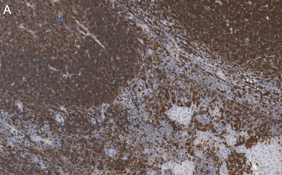
  • for immunofluorescence studies in tissue samples from human brain

Biochem/physiol Actions

Cofilin binds stoichiometrically to monomeric G-actin and actin protomers. Cofilin intercalates between longitudinally associated actin monomers within the filament. Under specific conditions, It cleaves the filaments and accelerates actin subunits dissociation from their ‘pointed′ ends. It is essential for viability and vital for many cellular processes involving actin remodeling, such as motility at the leading edge of cells, polarized cell growth, endocytosis, phagocytosis, cellular activation, cytokinesis etc. Cofilin activity is regulated through reversible phosphorylation and dephosphorylation. In phosphorylated form, it is inactive and unable to bond with actin. Phosphorylation of cofilin is regulated by at least four protein kinases: LIM Kinase-1, LIM Kinase-2, Testicular Kinase-1, and Testicular Kinase-2. S-nitrosylation of cofilin is involved in vascular endothelial growth factor (VEGF)-stimulated endothelial cytoskeleton remodeling and migration. Over-expression of cofilin suppresses growth and invasion of non-small cell lung cancer. Dysregulation of cofilin is associated with neurodegeneration.

Physical form

Solution in 0.01 M phosphate buffered saline, pH 7.4, containing 15 mM sodium azide.

Storage and Stability

For continuous use, store at 2-8 °C for up to one month. For prolonged storage, freeze in working aliquots at -20 °C. Repeated freezing and thawing, or storage in frost-free freezers, is not recommended. If slight turbidity occurs upon prolonged storage, clarify the solution by centrifugation before use. Working dilutions should be discarded if not used within 12 hours.

Disclaimer

Unless otherwise stated in our catalog or other company documentation accompanying the product(s), our products are intended for research use only and are not to be used for any other purpose, which includes but is not limited to, unauthorized commercial uses, in vitro diagnostic uses, ex vivo or in vivo therapeutic uses or any type of consumption or application to humans or animals.

Not finding the right product?  

Try our Product Selector Tool.

Storage Class Code

10 - Combustible liquids

WGK

WGK 1

Flash Point(F)

Not applicable

Flash Point(C)

Not applicable


Certificates of Analysis (COA)

Search for Certificates of Analysis (COA) by entering the products Lot/Batch Number. Lot and Batch Numbers can be found on a product’s label following the words ‘Lot’ or ‘Batch’.

Already Own This Product?

Find documentation for the products that you have recently purchased in the Document Library.

Visit the Document Library

Patrícia Schönhofen et al.
Mini reviews in medicinal chemistry, 14(5), 393-400 (2014-05-13)
Cofilin-1 protein, which main function is to regulate actin cytoskeleton dynamics, appears to be involved with many steps in the neurotoxicity processes found in neurodegenerative diseases such as Alzheimer's disease (AD), Parkinson's disease (PD) and Huntington's disease (HD). As the
Patrick J Hensley et al.
Urologic oncology, 37(9), 572-572 (2019-07-22)
Cisplatin-based neoadjuvant chemotherapy (NAC) for muscle-invasive urothelial carcinoma of the bladder confers only a modest survival advantage. Patients with pathologic progression on NAC have poor outcomes related to a delay in definitive surgical management. To characterize the value of epithelial-mesenchymal
Tasnim Rahman et al.
Journal of Alzheimer's disease : JAD, 42(4), 1443-1460 (2014-07-16)
Imaging of human brain as well as cellular and animal models has highlighted a role for the actin cytoskeleton in the development of cell pathology in Alzheimer's disease (AD). Rods and aggregates of the actin-associated protein cofilin are abundant in
Immune-complex level of cofilin-1 in sera is associated with cancer progression and poor prognosis in pancreatic cancer
Satoh M, et al.
Cancer Science, 108(4), 795-795 (2017)

Our team of scientists has experience in all areas of research including Life Science, Material Science, Chemical Synthesis, Chromatography, Analytical and many others.

Contact Technical Service