Skip to Content
Merck
All Photos(2)

Documents

C8487

Sigma-Aldrich

Anti-Caspase 3, Active antibody produced in rabbit

IgG fraction of antiserum, buffered aqueous solution

Synonym(s):

Activated Caspase 3 Antibody, Activated Caspase 3 Antibody - Anti-Caspase 3, Active antibody produced in rabbit, Anti-Apopain, Anti-CPP32, Anti-Yama

Sign Into View Organizational & Contract Pricing


About This Item

MDL number:
UNSPSC Code:
12352203
NACRES:
NA.41

biological source

rabbit

Quality Level

conjugate

unconjugated

antibody form

IgG fraction of antiserum

antibody product type

primary antibodies

clone

polyclonal

form

buffered aqueous solution

species reactivity

rat, bovine, human, pig, canine, mouse

packaging

antibody small pack of 25 μL

technique(s)

indirect immunofluorescence: 1:1,000 using human epitheloid carcinoma HeLa cell line, treated with staurosporine
microarray: suitable
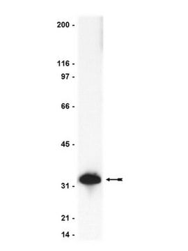
western blot: 1:500 using recombinant human caspase 3, active (Sigma Product No. C1224)

UniProt accession no.

shipped in

dry ice

storage temp.

−20°C

target post-translational modification

unmodified

Gene Information

human ... CASP3(836)
mouse ... Casp3(12367)
rat ... Casp3(25402)

General description

Caspase-3 (CASP3) belongs to the class of cysteine proteases. CASP3 is an executioner caspase, which is cleaved and activated with the help of caspase-8 and caspase-9 initiator caspases. CASP3 is mapped to human chromosome 4q35.1.
Caspases are a family of intracellular proteases that mediate cell death and the principal effectors of apoptosis. There are 14 different caspases have identified which can be grouped into three different subfamilies based on their substrate specificities. Caspase 3 is also known as CPP32, Yama, apopain. Among all the types, caspase 3, a cytosolic protein, found in cells as an inactive 32 kDa proenzyme.

Immunogen

Synthetic peptide corresponding to the cleavage site of human caspase 3 conjugated to KLH.

Application

Anti-Caspase 3, Active antibody has been used:
  • for western blotting of cytochrome c for caspase activation
  • as primary antibody in immunofluorescence staining of embryos and postnatal mice cryosections
  • in Western blot analysis of activated caspase 3
  • as a primary antibody in immunodetection of rat brain sections

Biochem/physiol Actions

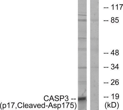
Caspase 3 is one of the key effectors of apoptosis. It acts downstream of caspase 9 in the apoptotic pathway. During apoptosis, it gets activated by proteolytic cleavage into the 17-19 kDa (p17, p18) and 12 kDa (p12) active subunits. Any alteraion in CASP-3 in mice, causes premature lethality. It has also shown that the deletion of gene affects brain development resulting of hyperplasias and disorganized cell deployement. Clinically it is related to Huntington disease (HD). Caspase 3 brings about cell death by activating CAD (caspase activated DNAse I), an endonuclease that causes degradation of chromosomal DNA.

Physical form

Supplied in 0.01 M phosphate buffered saline, pH 7.4, containing 15 mM sodium azide.

Storage and Stability

Store at –20 °C. For continuous use, the product may be storage, the product may be stored at 2-8 °C for up to one month. For prolonged storage, freeze in working aliquots at –20 °C. Repeated freezing and thawing, or storage in frost-free freezers, is not recommended. If slight turbidity occurs upon prolonged storage, clarify the solution by centrifugation before use. Working dilutions should be discarded if not used within 12 hours.

Disclaimer

Unless otherwise stated in our catalog or other company documentation accompanying the product(s), our products are intended for research use only and are not to be used for any other purpose, which includes but is not limited to, unauthorized commercial uses, in vitro diagnostic uses, ex vivo or in vivo therapeutic uses or any type of consumption or application to humans or animals.

Not finding the right product?  

Try our Product Selector Tool.

Storage Class Code

12 - Non Combustible Liquids

WGK

nwg

Flash Point(F)

Not applicable

Flash Point(C)

Not applicable


Certificates of Analysis (COA)

Search for Certificates of Analysis (COA) by entering the products Lot/Batch Number. Lot and Batch Numbers can be found on a product’s label following the words ‘Lot’ or ‘Batch’.

Already Own This Product?

Find documentation for the products that you have recently purchased in the Document Library.

Visit the Document Library

Annaïg Lan et al.
Apoptosis : an international journal on programmed cell death, 12(3), 573-591 (2006-12-30)
The human probiotic Propionibacterium freudenreichii kills colorectal adenocarcinoma cells through apoptosis in vitro via its metabolites, the short chain fatty acids (SCFA), acetate and propionate. However, the precise mechanisms, the kinetics of cellular events and the impact of environmental factors
Caspases: an apoptosis mediator
Palai T K and Mishra S R
Journal of Advanced Veterinary and Animal Research, 2(1) (2015)
Identification of SMURF1 as a possible target for 7q21. 3?22.1 amplification detected in a pancreatic cancer cell line by in?house array?based comparative genomic hybridization.
Suzuki A, et al.
Cancer Science, 99(5), 986-994 (2008)
Kalina Andreeva et al.
Ophthalmology and eye diseases, 6, 43-54 (2014-09-12)
Ischemia/reperfusion (IR) injury has been associated with several retinal pathologies, and a few genes/gene products have been linked to IR injury. However, the big picture of temporal changes, regarding the affected gene networks, pathways, and processes remains to be determined.
Ludger Leifeld et al.
The American journal of pathology, 168(3), 778-785 (2006-03-02)
Apoptosis is a key mechanism underlying fulminant hepatic failure. The role of gelsolin in such apoptotic pathways is not well understood because both pro-apoptotic and anti-apoptotic effects have been reported in vitro, depending on the cell type and in vitro

Our team of scientists has experience in all areas of research including Life Science, Material Science, Chemical Synthesis, Chromatography, Analytical and many others.

Contact Technical Service