Accéder au contenu
Merck
Toutes les photos(2)

Principaux documents

MABN69

Sigma-Aldrich

Anti-Caspr Antibody, clone K65/35

clone K65/35, from mouse

Synonyme(s) :

contactin associated protein 1, neurexin 4, neurexin-4, contactin-associated protein 1, Neurexin IV, Neurexin-4

Se connecterpour consulter vos tarifs contractuels et ceux de votre entreprise/organisme


About This Item

Code UNSPSC :
12352203
eCl@ss :
32160702
Nomenclature NACRES :
NA.41

Source biologique

mouse

Niveau de qualité

Forme d'anticorps

purified immunoglobulin

Type de produit anticorps

primary antibodies

Clone

K65/35, monoclonal

Espèces réactives

rat

Réactivité de l'espèce (prédite par homologie)

mouse (based on 100% sequence homology)

Technique(s)

immunohistochemistry: suitable
western blot: suitable

Isotype

IgG1κ

Numéro d'accès NCBI

Numéro d'accès UniProt

Conditions d'expédition

wet ice

Modification post-traductionnelle de la cible

unmodified

Informations sur le gène

Description générale

Contactin-associated protein 1(Caspr) is a single-pass type I membrane protein belonging to the neurexin family and is comprised of one F5/8 type C domain, one fibrinogen C-terminal domain, two EGF-like domains, and four laminin G-like domains. Caspr is well documented for its interaction with contactin, an interaction that is crucial to the transport of Caspr to the plasma membrane, the mylination of glial cells, and the generation of axoglial junctions. Studies also suggest that Caspr may play a regulatory role in the cell surface expression of contactin and its targeting to different axonal domains. Expression of Caspr is largely observed in the brain, mainly in CNS myelinated nerve fibers within paranodal axoglial junctions.

Immunogène

Recombinant protein corresponding to rat Caspr.

Application

Anti-Caspr Antibody, clone K65/35 is an antibody against Caspr for use in IH & WB.
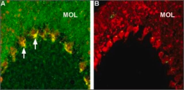
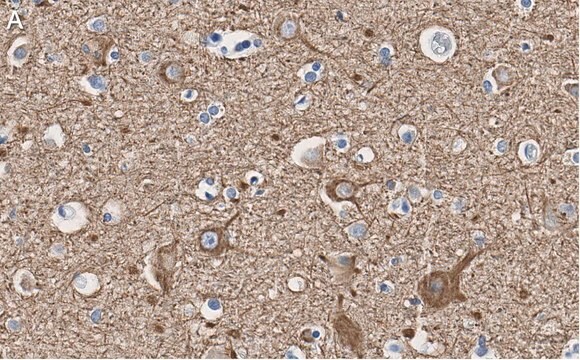
Immunohistochemistry Analysis: 1:300 dilution from a previous lot detected Caspr in rat cerebellum and rat hippocampus tissues.
Research Category
Neuroscience
Research Sub Category
Signaling Neuroscience

Qualité

Evaluated by Western Blot in rat brain membrane tissue lysate.

Western Blot Analysis: 0.5 µg/mL of this antibody detected Caspr on 10 µg of rat brain membrane tissue lysate.

Description de la cible

~ 220 kDa observed

Forme physique

Format: Purified
Protein G
Purified mouse monoclonal IgG1κ in buffer containing 0.1 M Tris-Glycine (pH 7.4), 150 mM NaCl with 0.05% sodium azide.

Stockage et stabilité

Stable for 1 year at 2-8°C from date of receipt.

Remarque sur l'analyse

Control
Rat brain membrane tissue lysate

Autres remarques

Concentration: Please refer to the Certificate of Analysis for the lot-specific concentration.

Clause de non-responsabilité

Unless otherwise stated in our catalog or other company documentation accompanying the product(s), our products are intended for research use only and are not to be used for any other purpose, which includes but is not limited to, unauthorized commercial uses, in vitro diagnostic uses, ex vivo or in vivo therapeutic uses or any type of consumption or application to humans or animals.

Vous ne trouvez pas le bon produit ?  

Essayez notre Outil de sélection de produits.

Code de la classe de stockage

12 - Non Combustible Liquids

Classe de danger pour l'eau (WGK)

WGK 1

Point d'éclair (°F)

Not applicable

Point d'éclair (°C)

Not applicable


Certificats d'analyse (COA)

Recherchez un Certificats d'analyse (COA) en saisissant le numéro de lot du produit. Les numéros de lot figurent sur l'étiquette du produit après les mots "Lot" ou "Batch".

Déjà en possession de ce produit ?

Retrouvez la documentation relative aux produits que vous avez récemment achetés dans la Bibliothèque de documents.

Consulter la Bibliothèque de documents

Annika Balraj et al.
Developmental neurobiology, 82(4), 308-325 (2022-04-12)
Retinal ganglion cells generate a pattern of action potentials to communicate visual information from the retina to cortical areas. Myelin, an insulating sheath, wraps axonal segments to facilitate signal propagation and when deficient, can impair visual function. Optic nerve development
Jacob C A Edvinsson et al.
The journal of headache and pain, 23(1), 26-26 (2022-02-19)
5-Hydroxytryptamine (5-HT) receptors 1B, 1D and 1F have key roles in migraine pharmacotherapy. Selective agonists targeting these receptors, such as triptans and ditans, are effective in aborting acute migraine attacks and inhibit the in vivo release of calcitonin gene-related peptide
Anirban Paul et al.
Cell, 171(3), 522-539 (2017-09-26)
Understanding the organizational logic of neural circuits requires deciphering the biological basis of neuronal diversity and identity, but there is no consensus on how neuron types should be defined. We analyzed single-cell transcriptomes of a set of anatomically and physiologically characterized
Aníbal Sánchez-de la Torre et al.
Cell death & disease, 13(7), 585-585 (2022-07-08)
Cannabinoids are known to modulate oligodendrogenesis and developmental CNS myelination. However, the cell-autonomous action of these compounds on oligodendroglial cells in vivo, and the molecular mechanisms underlying these effects have not yet been studied. Here, by using oligodendroglial precursor cell
Ana Palma et al.
Frontiers in cell and developmental biology, 10, 841548-841548 (2022-04-05)
Abnormalities in myelination are associated to behavioral and cognitive dysfunction in neurodevelopmental psychiatric disorders. Thus, therapies to promote or accelerate myelination could potentially ameliorate symptoms in autism. Clemastine, a histamine H1 antagonist with anticholinergic properties against muscarinic M1 receptor, is

Notre équipe de scientifiques dispose d'une expérience dans tous les secteurs de la recherche, notamment en sciences de la vie, science des matériaux, synthèse chimique, chromatographie, analyse et dans de nombreux autres domaines..

Contacter notre Service technique