AB3768
Anti-Rad54L Antibody
Chemicon®, from rabbit
About This Item
Produits recommandés
Source biologique
rabbit
Niveau de qualité
Forme d'anticorps
saturated ammonium sulfate (SAS) precipitated
Type de produit anticorps
primary antibodies
Clone
polyclonal
Produit purifié par
affinity chromatography
Espèces réactives
human, mouse
Fabricant/nom de marque
Chemicon®
Technique(s)
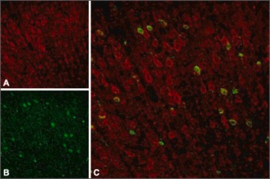
immunocytochemistry: suitable
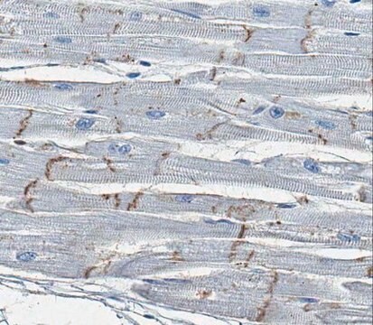
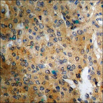
immunohistochemistry: suitable
Numéro d'accès NCBI
Numéro d'accès UniProt
Conditions d'expédition
dry ice
Modification post-traductionnelle de la cible
unmodified
Informations sur le gène
human ... RAD54L(8438)
Spécificité
Immunogène
Application
Immunocytochemistry: 1:200-1:1,000.
Optimal working dilutions must be determined by the end user.SUGGESTED PROTOCOL FOR IMMUNOHISTOCHEMISTRY
1. Dissect tissues and freeze on dry ice
2. Cut on a cryostat - 10um slices
3. Dry slides at room temperature for 30-90 min
4. Fix with cold Acetone/Methanol (50/50) 2 min - then either process further or store at -20°C . Alternative is fixation with 4% PFA and treatment with a trypsin solution. (0.05% trypsin , incubation 10 minutes at 37°C, followed by 3 washes with PBS)
5. Let air dry
6. PBS 5min
7. Incubate in 50mM ammonium chloride 30 min
8. PBS 5min
9. Blocking serum 30-45min
10. Primary antibody 90min - dilution 1:100 - 1:600
11. Wash 3 times for 5 min
12. Secondary fluorescent conjug. antibody 30min
13. Wash 3 times for 5 min and mount
Epigenetics & Nuclear Function
Cell Cycle, DNA Replication & Repair
Forme physique
PREPARATION AND USE: To reconstitute the antibody, centrifuge the antibody vial at moderate speed (5,000 rpm) for 5 minutes to pellet the precipitated antibody product. Carefully remove the ammonium sulfate/PBS buffer solution and discard. It is not necessary to remove all of the ammonium sulfate/PBS solution: 10 μL of residual ammonium sulfate solution will not effect the resuspension of the antibody. Do not let the protein pellet dry, as severe loss of antibody reactivity can occur.
Resuspend the antibody pellet in any suitable biological buffer, standard PBS or TBS (pH 7.3-7.5) are typical. Volumes required are not critical but it is suggested that the final antibody concentration be between 0.1 mg/mL and 1.0 mg/mL. For example, to achieve a 1 mg/mL concentration with 50 μg of precipitated antibody, the amount of buffer needed would be 50 μL.
Carefully add the liquid buffer to the pellet. DO NOT VORTEX. Mix by gentle stirring with a wide pipet tip or gentle finger-tapping. Let the precipitated antibody rehydrate for 1 hour at 4-25C° prior to use. Small particles of precipitated antibody that fail to resuspend are normal. Vials are overfilled to compensate for any losses.
Stockage et stabilité
Informations légales
Clause de non-responsabilité
Vous ne trouvez pas le bon produit ?
Essayez notre Outil de sélection de produits.
Code de la classe de stockage
12 - Non Combustible Liquids
Classe de danger pour l'eau (WGK)
WGK 2
Point d'éclair (°F)
Not applicable
Point d'éclair (°C)
Not applicable
Certificats d'analyse (COA)
Recherchez un Certificats d'analyse (COA) en saisissant le numéro de lot du produit. Les numéros de lot figurent sur l'étiquette du produit après les mots "Lot" ou "Batch".
Déjà en possession de ce produit ?
Retrouvez la documentation relative aux produits que vous avez récemment achetés dans la Bibliothèque de documents.
Notre équipe de scientifiques dispose d'une expérience dans tous les secteurs de la recherche, notamment en sciences de la vie, science des matériaux, synthèse chimique, chromatographie, analyse et dans de nombreux autres domaines..
Contacter notre Service technique


