Skip to Content
Merck
All Photos(1)

Key Documents

A5964

Sigma-Aldrich

Monoclonal Anti-Phosphotyrosine–Peroxidase antibody produced in mouse

clone PT-66, purified immunoglobulin, lyophilized powder

Synonym(s):

Monoclonal Anti-Phosphotyrosine, Phospho-Tyr, Phospho-tyrosine, p-Tyr

Sign Into View Organizational & Contract Pricing


About This Item

UNSPSC Code:
12352203
NACRES:
NA.43

biological source

mouse

Quality Level

conjugate

peroxidase conjugate

antibody form

purified immunoglobulin

antibody product type

primary antibodies

clone

PT-66, monoclonal

form

lyophilized powder

packaging

vial of 0.2 mL conjugate

technique(s)

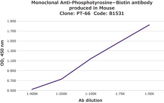
direct ELISA: 1:60,000 using Phosphotyrosine-BSA
dot blot: 1:40,000-1:200,000 using phosphotyrosine-BSA using chromogenic and chemiluminescent substrates, respectively

isotype

IgG1

storage temp.

2-8°C

target post-translational modification

unmodified

Looking for similar products? Visit Product Comparison Guide

General description

As determined by ELISA and competitive ELISA, the antibody reacts specifically with phosphorylated tyrosine, both as free amino acid or conjugated to carriers such as BSA or KLH. No cross-reactivity is observed with non-phosphorylated tyrosine, phosphothreonine, phosphoserine, AMP or ATP.
Monoclonal Anti-Phosphotyrosine (mouse IgG1 isotype) is derived from the PT-66 hybridoma produced by the fusion of mouse myeloma cells and splenocytes from BALB/c mice immunized with a phosphotyrosine-BSA conjugate.

Specificity

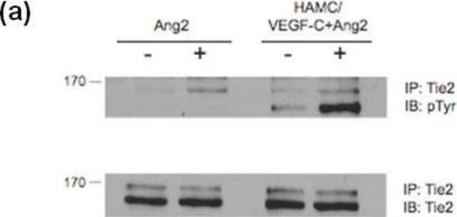
Mouse anti-phosphotyrosine-peroxidase antibody reacts specifically with phosphorylated tyrosine coupled to BSA. The product has shown no reactivity for other phosphorylated amino acids like phosphothreonine and phosphoserine.

Immunogen

phosphotyrosine conjugated to BSA

Application

Monoclonal Anti-Phosphotyrosine-Peroxidase antibody has been used in
  • enzyme linked immunosorbent assay (ELISA)
  • dot blot
  • Chemiluminescence dot blot
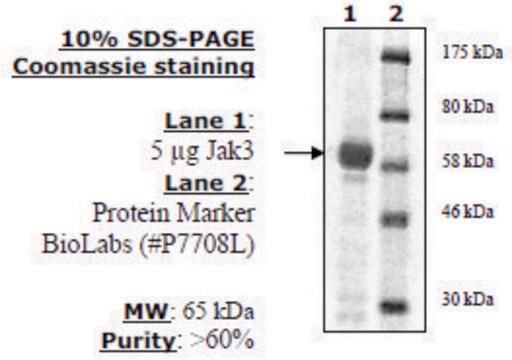
  • kinase assay

Biochem/physiol Actions

Reversible phosphorylation of proteins is an important post-translational modification that plays a regulatory role in the expression of most proteins in the cells. Reversible phosphorylation at multiple serine, tyrosine and threonine residues mediates numerous signalling pathways in both prokaryotic and eukaryotic cells . Cellular proteins with phosphorylated tyrosine increase many fold by the activation of tyrosine kinases. Most mitogenic receptor systems such as EGF, PDGF, insulin receptors contain serine/threonine/tyrosine kinase domains that undergo autophosphorylation when receptors bind to the respective ligands. Monoclonal anti-phosphotyrosine–peroxidase antibody can be used in kinase assay to visualize the fraction of phosphorylated substrate.

Physical form

Lyophilized from a solution containing 1% bovine serum albumin and 0.05% MIT in 0.01 M sodium phosphate buffered saline.

Disclaimer

Unless otherwise stated in our catalog or other company documentation accompanying the product(s), our products are intended for research use only and are not to be used for any other purpose, which includes but is not limited to, unauthorized commercial uses, in vitro diagnostic uses, ex vivo or in vivo therapeutic uses or any type of consumption or application to humans or animals.

Not finding the right product?  

Try our Product Selector Tool.

Storage Class Code

13 - Non Combustible Solids

WGK

WGK 3

Flash Point(F)

Not applicable

Flash Point(C)

Not applicable


Certificates of Analysis (COA)

Search for Certificates of Analysis (COA) by entering the products Lot/Batch Number. Lot and Batch Numbers can be found on a product’s label following the words ‘Lot’ or ‘Batch’.

Already Own This Product?

Find documentation for the products that you have recently purchased in the Document Library.

Visit the Document Library

Bobbi Xayarath et al.
Journal of bacteriology, 189(9), 3369-3381 (2007-02-27)
Extracellular polysaccharides of many bacteria are synthesized by the Wzy polymerase-dependent mechanism, where long-chain polymers are assembled from undecaprenyl-phosphate-linked repeat units on the outer face of the cytoplasmic membrane. In gram-positive bacteria, Wzy-dependent capsules remain largely cell associated via membrane
Identification and biophysical assessment of the molecular recognition mechanisms between the human haemopoietic cell kinase Src homology domain 3 and ALG-2-interacting protein X
Shi X, et al.
The Biochemical Journal, 431(1), 93-102 (2010)
Yuko Naito et al.
Molecular and cellular biology, 27(8), 3008-3022 (2007-02-14)
Sialic acid (Sia) is a family of acidic nine-carbon sugars that occupies the nonreducing terminus of glycan chains. Diversity of Sia is achieved by variation in the linkage to the underlying sugar and modification of the Sia molecule. Here we
Xiaoli Shi et al.
The Biochemical journal, 431(1), 93-102 (2010-07-31)
SFKs (Src family kinases) are central regulators of many signalling pathways. Their functions are tightly regulated through SH (Src homology) domain-mediated protein-protein interactions. A yeast two-hybrid screen using SH3 domains as bait identified Alix [ALG-2 (apoptosis-linked gene 2)-interacting protein X]
Dan Su et al.
Evidence-based complementary and alternative medicine : eCAM, 2019, 4982062-4982062 (2019-05-17)
The Janus kinases (JAKs) consist of four similar tyrosine kinases and function as key hubs in the signaling pathways that are implicated in both innate and adaptive immunity. Among the four members, JAK3 is probably the more attractive target for

Our team of scientists has experience in all areas of research including Life Science, Material Science, Chemical Synthesis, Chromatography, Analytical and many others.

Contact Technical Service