Skip to Content
Merck
All Photos(2)

Documents

AB5164

Sigma-Aldrich

Anti-Muscarinic Acetylcholine Receptor m1 Antibody

Chemicon®, from rabbit

Sign Into View Organizational & Contract Pricing


About This Item

UNSPSC Code:
12352203
eCl@ss:
32160702
NACRES:
NA.43

biological source

rabbit

Quality Level

antibody form

affinity purified immunoglobulin

antibody product type

primary antibodies

clone

polyclonal

purified by

affinity chromatography

species reactivity

rat, mouse

manufacturer/tradename

Chemicon®

technique(s)

immunohistochemistry: suitable
western blot: suitable

NCBI accession no.

UniProt accession no.

shipped in

wet ice

target post-translational modification

unmodified

Gene Information

human ... CHRM1(1128)

Specificity

Recognizes a full length m1 protein. It has exhibited no cross reactivity with other muscarinic proteins tested so far.

Immunogen

GST fusion protein and part of i3 intercellular loop of human m1 muscarinic acetylcholine receptor (amino acids 227-353) (Accession P11229).

Application

Research Category
Neuroscience
Research Sub Category
Neurotransmitters & Receptors
This Anti-Muscarinic Acetylcholine Receptor m1 Antibody is validated for use in IH, WB for the detection of Muscarinic Acetylcholine Receptor m1.
Western blot: 1:200 using ECL on rat brain membranes with proteinase inhibitors. M1 is approximately 60-80kDa in size, some preparations yield 125kDa dimers (photo). Heating samples to 80-90C rather than fulling boiling and adjusting sample pH to neutral can help in preventing dimer formation.

Dilutions should be made using a carrier protein such as BSA (1-3%)

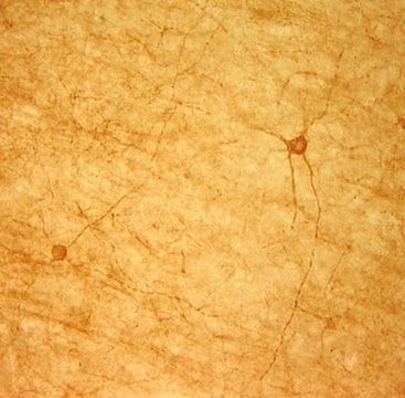
Immunohistochemistry: rat frozen sections. 1:50-1:200, internal epitope; use triton X-100 in blocking buffer only; dilute primary antibody in PBS or TBS with 0.5%-1% BSA or NGS only.


Optimal working dilutions must be determined by the end user.

Physical form

Affinity purified immunoglobulin. Lyophilized from phosphate buffered saline, pH 7.4, containing 1% BSA and 0.05% sodium azide as a preservative. Reconstitute with 200 μL of sterile deionized water. Centrifuge antibody preparation before use (10,000 x g for 5 min).

Storage and Stability

Maintain lyophilized material at -20°C for up to 12 months after date of receipt. After reconstitution maintain at -20°C in undiluted aliquots for up to 6 months. Avoid repeated freeze/thaw cycles.

Analysis Note

Control
Included free of charge with the antibody is 60 μg of control fusion protein (lyophilized powder). The stock solution of the fusion protein can be made up using 100 μL of PBS. For positive control, in Western blot using 10 ng of protein per minigel lane. For negative control, preincubate 3 μg of fusion protein with 1 μg of antibody for one hour at room temperature. Optimal concentrations must be determined by the end user.

Other Notes

Concentration: Please refer to the Certificate of Analysis for the lot-specific concentration.

Legal Information

CHEMICON is a registered trademark of Merck KGaA, Darmstadt, Germany

Disclaimer

Unless otherwise stated in our catalog or other company documentation accompanying the product(s), our products are intended for research use only and are not to be used for any other purpose, which includes but is not limited to, unauthorized commercial uses, in vitro diagnostic uses, ex vivo or in vivo therapeutic uses or any type of consumption or application to humans or animals.

Not finding the right product?  

Try our Product Selector Tool.

Hazard Statements

Precautionary Statements

Hazard Classifications

Aquatic Chronic 3

Storage Class Code

11 - Combustible Solids

WGK

WGK 3


Certificates of Analysis (COA)

Search for Certificates of Analysis (COA) by entering the products Lot/Batch Number. Lot and Batch Numbers can be found on a product’s label following the words ‘Lot’ or ‘Batch’.

Already Own This Product?

Find documentation for the products that you have recently purchased in the Document Library.

Visit the Document Library

M Takamori et al.
European journal of neurology, 14(11), 1230-1235 (2007-09-04)
The Lambert-Eaton myasthenic syndrome (LEMS), often associated with small-cell lung carcinoma (SCLC), is a disorder of acetylcholine (ACh) release from motor nerve terminals. In most patients, it is caused by autoantibodies against the P/Q-type voltage-gated calcium channels (VGCC) that trigger
Marci L Smith et al.
Molecular vision, 20, 1328-1356 (2014-10-30)
The α7 nicotinic acetylcholine receptor (nAChR) is widely expressed in the nervous system, including in the inner retinal neurons in all species studied to date. Although reductions in the expression of α7 nAChRs are thought to contribute to the memory
Gitte Jositsch et al.
Naunyn-Schmiedeberg's archives of pharmacology, 379(4), 389-395 (2008-11-01)
Acetylcholine (ACh) is a major regulator of visceral function exerting pharmacologically relevant effects upon smooth muscle tone and epithelial function via five types of muscarinic receptors (M1R-M5R). In this paper, we assessed the specificity of muscarinic receptor (MR) antibodies in
A M Harrington et al.
Neurogastroenterology and motility : the official journal of the European Gastrointestinal Motility Society, 22(9), 999-1008 (2010-02-12)
Muscarinic acetylcholine receptors (MR) are involved in multiple intestinal reflexes. The cellular localization of subtypes of MRs within enteric circuits mediating muscle and mucosal reflexes remains to be demonstrated. This study aimed to localize the three functionally significant subtypes of
Chase M Carver et al.
Frontiers in neuroscience, 15, 681144-681144 (2021-09-08)
We investigated the contribution of excitatory transient receptor potential canonical (TRPC) cation channels to posttraumatic hyperexcitability in the brain 7 days following controlled cortical impact model of traumatic brain injury (TBI) to the parietal cortex in male adult mice. We

Our team of scientists has experience in all areas of research including Life Science, Material Science, Chemical Synthesis, Chromatography, Analytical and many others.

Contact Technical Service