Accéder au contenu
Merck
Toutes les photos(7)

Documents

HPA014907

Sigma-Aldrich

Anti-PDZK1IP1 antibody produced in rabbit

enhanced validation

Prestige Antibodies® Powered by Atlas Antibodies, affinity isolated antibody, buffered aqueous glycerol solution

Synonyme(s) :

Anti-17 kDa membrane-associated protein, Anti-PDZK1-interacting protein 1, Anti-Protein DD96

Se connecterpour consulter vos tarifs contractuels et ceux de votre entreprise/organisme


About This Item

Code UNSPSC :
12352203
Numéro HPA (Human Protein Atlas):
Nomenclature NACRES :
NA.41

Source biologique

rabbit

Conjugué

unconjugated

Forme d'anticorps

affinity isolated antibody

Type de produit anticorps

primary antibodies

Clone

polyclonal

Gamme de produits

Prestige Antibodies® Powered by Atlas Antibodies

Forme

buffered aqueous glycerol solution

Espèces réactives

human

Validation améliorée

orthogonal RNAseq
recombinant expression
Learn more about Antibody Enhanced Validation

Technique(s)

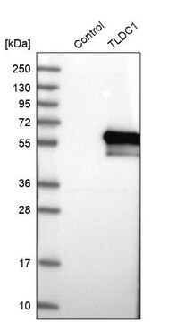
immunoblotting: 0.04-0.4 μg/mL
immunofluorescence: 0.25-2 μg/mL
immunohistochemistry: 1:200-1:500

Séquence immunogène

QEEPEPAHMILTVGNKADGVLVGTDGRYSSMAASFRSSEHENAYENVPEEEGKVRST

Numéro d'accès UniProt

Conditions d'expédition

wet ice

Température de stockage

−20°C

Modification post-traductionnelle de la cible

unmodified

Informations sur le gène

Description générale

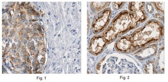
PDZK1IP1 (PDZK1 interacting protein 1) is a non-glycosylated membrane protein, which resides in the plasma membrane and Golgi bodies. It has a hydrophobic N-terminal of 13 residues, making the PDZ-binding domain, and two transmembrane regions. The molecular weight of this protein is 17kDa, and is expressed significantly only in normal kidney, in the proximal tubular epithelial cells.

Immunogène

PDZK1-interacting protein 1 recombinant protein epitope signature tag (PrEST)

Application

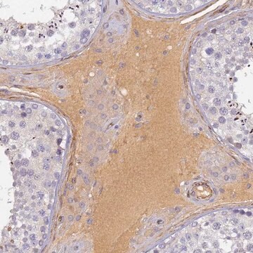
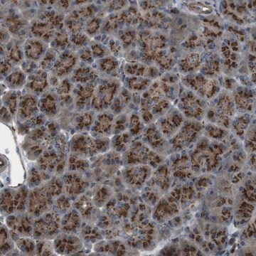
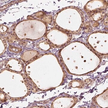
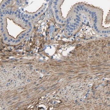
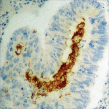
All Prestige Antibodies Powered by Atlas Antibodies are developed and validated by the Human Protein Atlas (HPA) project and as a result, are supported by the most extensive characterization in the industry.

The Human Protein Atlas project can be subdivided into three efforts: Human Tissue Atlas, Cancer Atlas, and Human Cell Atlas. The antibodies that have been generated in support of the Tissue and Cancer Atlas projects have been tested by immunohistochemistry against hundreds of normal and disease tissues and through the recent efforts of the Human Cell Atlas project, many have been characterized by immunofluorescence to map the human proteome not only at the tissue level but now at the subcellular level. These images and the collection of this vast data set can be viewed on the Human Protein Atlas (HPA) site by clicking on the Image Gallery link. We also provide Prestige Antibodies® protocols and other useful information.

Actions biochimiques/physiologiques

PDZK1IP1 (PDZK1 interacting protein 1) acts as an anchor for PDZK1 (PDZ domain containing 1) protein, and in renal proximal tubules interacts with NaPi-IIa/PDZK1 comlex. In human cancer cells, it facilitates the Na-dependent transport of mannose and glucose, by interacting with SGLT transporters. Amplification of mRNA leads to its elevated levels in multiple human tumors, and this relates with tumor progression, as seen in breast, ovarian and prostate cancer. Overexpression of this gene in tumors is associated with good prognosis. This protein induces PI3K/AKT pathway, which leads to prevention of Myc-induced apoptosis. Its PDZ-binding domain is responsible for its capacity to increase reactive oxygen species (ROS), which in turn is implicated in tumorigenesis.

Caractéristiques et avantages

Prestige Antibodies® are highly characterized and extensively validated antibodies with the added benefit of all available characterization data for each target being accessible via the Human Protein Atlas portal linked just below the product name at the top of this page. The uniqueness and low cross-reactivity of the Prestige Antibodies® to other proteins are due to a thorough selection of antigen regions, affinity purification, and stringent selection. Prestige antigen controls are available for every corresponding Prestige Antibody and can be found in the linkage section.

Every Prestige Antibody is tested in the following ways:
  • IHC tissue array of 44 normal human tissues and 20 of the most common cancer type tissues.
  • Protein array of 364 human recombinant protein fragments.

Liaison

Corresponding Antigen APREST72299

Forme physique

Solution in phosphate-buffered saline, pH 7.2, containing 40% glycerol and 0.02% sodium azide

Informations légales

Prestige Antibodies is a registered trademark of Merck KGaA, Darmstadt, Germany

Clause de non-responsabilité

Unless otherwise stated in our catalog or other company documentation accompanying the product(s), our products are intended for research use only and are not to be used for any other purpose, which includes but is not limited to, unauthorized commercial uses, in vitro diagnostic uses, ex vivo or in vivo therapeutic uses or any type of consumption or application to humans or animals.

Not finding the right product?  

Try our Outil de sélection de produits.

Code de la classe de stockage

10 - Combustible liquids

Classe de danger pour l'eau (WGK)

WGK 1

Point d'éclair (°F)

Not applicable

Point d'éclair (°C)

Not applicable

Équipement de protection individuelle

Eyeshields, Gloves, multi-purpose combination respirator cartridge (US)


Certificats d'analyse (COA)

Recherchez un Certificats d'analyse (COA) en saisissant le numéro de lot du produit. Les numéros de lot figurent sur l'étiquette du produit après les mots "Lot" ou "Batch".

Déjà en possession de ce produit ?

Retrouvez la documentation relative aux produits que vous avez récemment achetés dans la Bibliothèque de documents.

Consulter la Bibliothèque de documents

Lucile Massenet-Regad et al.
iScience, 26(12), 108367-108367 (2023-11-29)
Cellular crosstalk in the tumor microenvironment (TME) is still largely uncharacterized, while it plays an essential role in shaping immunosuppression or anti-tumor response. Large-scale analyses are needed to better decipher cell-cell communication in cancer. In this work, we used original
Royce W Zhou et al.
Nature communications, 13(1), 6041-6041 (2022-10-18)
Tumors exhibit enhancer reprogramming compared to normal tissue. The etiology is largely attributed to cell-intrinsic genomic alterations. Here, using freshly resected primary CRC tumors and patient-matched adjacent normal colon, we find divergent epigenetic landscapes between CRC tumors and cell lines.
Maria V Guijarro et al.
Carcinogenesis, 28(12), 2443-2450 (2007-08-07)
MAP17 is a non-glycosylated membrane-associated protein that has been shown to be over-expressed in human carcinomas, suggesting a possible role of this protein in tumorigenesis. However, very little is known about the molecular mechanism mediating the possible tumor promoting properties
Maria V Guijarro et al.
Carcinogenesis, 28(10), 2096-2104 (2007-06-06)
Tumorigenesis occurs when the mechanisms involved in the control of tissue homeostasis are disrupted and cells stop responding to physiological signals. Therefore, genes capable of desensitizing tumoral cells from physiological signals may provide a selective advantage within the tumoral mass
Marco Perez et al.
PloS one, 8(2), e56169-e56169 (2013-02-19)
MAP17 is a membrane-associated protein that is overexpressed in human tumors. Because the expression of MAP17 increases reactive oxygen species (ROS) generation through SGLT1 in cancer cells, in the present work, we investigated whether MAP17 and/or SGLT1 might be markers

Notre équipe de scientifiques dispose d'une expérience dans tous les secteurs de la recherche, notamment en sciences de la vie, science des matériaux, synthèse chimique, chromatographie, analyse et dans de nombreux autres domaines..

Contacter notre Service technique