Accéder au contenu
Merck
Toutes les photos(7)

Documents

HPA007973

Sigma-Aldrich

Anti-S100A4 antibody produced in rabbit

enhanced validation

Prestige Antibodies® Powered by Atlas Antibodies, affinity isolated antibody, buffered aqueous glycerol solution

Synonyme(s) :

Anti-Calvasculin antibody produced in rabbit, Anti-Metastasin antibody produced in rabbit, Anti-Placental calcium-binding protein antibody produced in rabbit, Anti-Protein Mts1 antibody produced in rabbit, Anti-Protein S100-A4 antibody produced in rabbit, Anti-S100 calcium-binding protein A4 antibody produced in rabbit

Se connecterpour consulter vos tarifs contractuels et ceux de votre entreprise/organisme


About This Item

Code UNSPSC :
12352203
Numéro HPA (Human Protein Atlas):
Nomenclature NACRES :
NA.41

Source biologique

rabbit

Conjugué

unconjugated

Forme d'anticorps

affinity isolated antibody

Type de produit anticorps

primary antibodies

Clone

polyclonal

Gamme de produits

Prestige Antibodies® Powered by Atlas Antibodies

Forme

buffered aqueous glycerol solution

Espèces réactives

mouse, human, rat

Validation améliorée

RNAi knockdown
recombinant expression
orthogonal RNAseq
Learn more about Antibody Enhanced Validation

Technique(s)

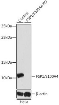
immunoblotting: 0.04-0.4 μg/mL
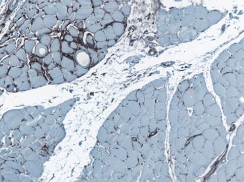
immunohistochemistry: 1:1000-1:2500

Séquence immunogène

MACPLEKALDVMVSTFHKYSGKEGDKFKLNKSELKELLTRELPSFLGKRTDEAAFQKLMSNLDSNRDNEVDFQEYCVFLSCIAMMCNEFFEGFPDKQPRKK

Numéro d'accès UniProt

Conditions d'expédition

wet ice

Température de stockage

−20°C

Modification post-traductionnelle de la cible

unmodified

Informations sur le gène

human ... S100A4(6275)

Description générale

Rabbit polyclonal anti-S100A4 antibody reacts with human S100 calcium-binding protein A4.
S100 calcium-binding protein A4 (S100A4) is a member of the S100 family of calcium binding proteins. S100A4 has been identified as a major prometastatic protein that is upregulated in invasive tumors wherein it contributes to tumor metastasis. S100A4 is a useful target for studies on tumor metastasis and potential therapeutic development.

Immunogène

Protein S100-A4 recombinant protein epitope signature tag (PrEST)

Application

All Prestige Antibodies Powered by Atlas Antibodies are developed and validated by the Human Protein Atlas (HPA) project and as a result, are supported by the most extensive characterization in the industry.

The Human Protein Atlas project can be subdivided into three efforts: Human Tissue Atlas, Cancer Atlas, and Human Cell Atlas. The antibodies that have been generated in support of the Tissue and Cancer Atlas projects have been tested by immunohistochemistry against hundreds of normal and disease tissues and through the recent efforts of the Human Cell Atlas project, many have been characterized by immunofluorescence to map the human proteome not only at the tissue level but now at the subcellular level. These images and the collection of this vast data set can be viewed on the Human Protein Atlas (HPA) site by clicking on the Image Gallery link. We also provide Prestige Antibodies® protocols and other useful information.
Rabbit polyclonal anti-S100A4 antibody is used to tag S100 calcium-binding protein A4 for detection and quantitation by immunocytochemical and immunohistochemical (IHC) techniques. It is used as a probe to determine the presence and roles of S100 calcium-binding protein A4 in calcium mediated cell signaling and tumor metastasis.

Caractéristiques et avantages

Prestige Antibodies® are highly characterized and extensively validated antibodies with the added benefit of all available characterization data for each target being accessible via the Human Protein Atlas portal linked just below the product name at the top of this page. The uniqueness and low cross-reactivity of the Prestige Antibodies® to other proteins are due to a thorough selection of antigen regions, affinity purification, and stringent selection. Prestige antigen controls are available for every corresponding Prestige Antibody and can be found in the linkage section.

Every Prestige Antibody is tested in the following ways:
  • IHC tissue array of 44 normal human tissues and 20 of the most common cancer type tissues.
  • Protein array of 364 human recombinant protein fragments.

Liaison

Corresponding Antigen APREST70188

Forme physique

Solution in phosphate-buffered saline, pH 7.2, containing 40% glycerol and 0.02% sodium azide

Informations légales

Prestige Antibodies is a registered trademark of Merck KGaA, Darmstadt, Germany

Clause de non-responsabilité

Unless otherwise stated in our catalog or other company documentation accompanying the product(s), our products are intended for research use only and are not to be used for any other purpose, which includes but is not limited to, unauthorized commercial uses, in vitro diagnostic uses, ex vivo or in vivo therapeutic uses or any type of consumption or application to humans or animals.

Not finding the right product?  

Try our Outil de sélection de produits.

Code de la classe de stockage

10 - Combustible liquids

Classe de danger pour l'eau (WGK)

WGK 1

Point d'éclair (°F)

Not applicable

Point d'éclair (°C)

Not applicable

Équipement de protection individuelle

Eyeshields, Gloves, multi-purpose combination respirator cartridge (US)


Certificats d'analyse (COA)

Recherchez un Certificats d'analyse (COA) en saisissant le numéro de lot du produit. Les numéros de lot figurent sur l'étiquette du produit après les mots "Lot" ou "Batch".

Déjà en possession de ce produit ?

Retrouvez la documentation relative aux produits que vous avez récemment achetés dans la Bibliothèque de documents.

Consulter la Bibliothèque de documents

Johan A den Boon et al.
Proceedings of the National Academy of Sciences of the United States of America, 112(25), E3255-E3264 (2015-06-10)
To study the multistep process of cervical cancer development, we analyzed 128 frozen cervical samples spanning normalcy, increasingly severe cervical intraepithelial neoplasia (CIN1- CIN3), and cervical cancer (CxCa) from multiple perspectives, revealing a cascade of progressive changes. Compared with normal
Protein atlas of fibroblast specific protein 1 (FSP1)/S100A4.
Fendt, et al.
Histology and Histopathology, 18621-18621 (2023)
Xingbo Xu et al.
The Journal of biological chemistry, 290(27), 16653-16664 (2015-05-15)
Endothelial to mesenchymal transition (EndMT) was originally described in heart development where the endocardial endothelial cells that line the atrioventricular canal undergo an EndMT to form the endocardial mesenchymal cushion that later gives rise to the septum and mitral and
Johanna W Hellinger et al.
Frontiers in oncology, 9, 1074-1074 (2019-11-12)
Background and Objective: Matricellular proteins modulate the micro environment of tumors and are recognized to contribute to tumor cell invasion and dissemination. The cysteine-rich angiogenic inducer 61 (CYR61) is upregulated in mesenchymal transformed and invasive breast cancer cells. CYR61 correlates
Laura Lopez-Sanz et al.
Clinical and translational medicine, 11(7), e463-e463 (2021-07-30)
Abdominal aortic aneurysm (AAA), a degenerative vascular pathology characterized by permanent dilation of the aorta, is considered a chronic inflammatory disease involving innate/adaptive immunity. However, the functional role of antibody-dependent immune response against antigens present in the damaged vessel remains

Notre équipe de scientifiques dispose d'une expérience dans tous les secteurs de la recherche, notamment en sciences de la vie, science des matériaux, synthèse chimique, chromatographie, analyse et dans de nombreux autres domaines..

Contacter notre Service technique