MDM2 is an E3 ubiquitin ligase that is most widely studied for its role in regulating the p53 tumor suppressor. MDM2 antagonizes the transcriptional activity of p53 in two main ways. The N-terminal of MDM2 interacts and disables the transactivation domain of p53. In addition, the C-terminal E3 ligase region of MDM2 can attach monoubiquitin to p53 resulting in the transport of p53 from the nucleus to the cytoplasm, thereby preventing the interaction of p53 with target DNA sequences. In addition, MDM2 may also attach polyubiquitin chains to p53, resulting in the degradation of p53 by the proteosome. MDM2 may produce these effects in cooperation with the Mdmx protein. Overall, MDM2 inhibits the ability of p53 to induce cell cycle arrest or apoptosis, and may contribute to oncogenesis.
Application
Anti-MDM2 Antibody, clone 2A10 is a Mouse Monoclonal Antibody for detection of MDM2 also known as E3 ubiquitin-protein ligase Mdm2 or Hdm2 & has been validated in WB & IP.
Immunoprecipitation Analysis: A representative lot was used by an independent laboratory in human lymphoblastoid cell line (NL553) treated with NCS. (Khosravi, R., et al. (1999). PNAS. 96(26):14973–14977.)
Quality
Evaluated by Western Blot in A-549 cell lysate.
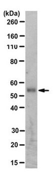
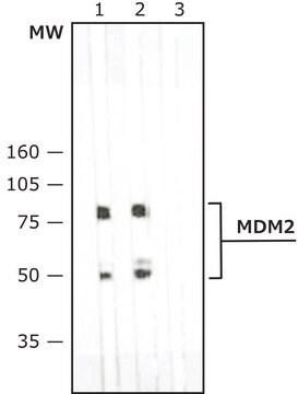
Western Blot Analysis: 0.5 µg/mL of this antibody detected MDM2 on 10 µg of A-549 cell lysate.
Target description
~90 kDa observed. The calculated molecular weight is 55 kDa, however MDM2 has been shown as a ~90 kDa band in western blots (Erhardt, P., et al. (1997). The Journal of Biological Chemistry. 272:15049-15052.). Uniprot describes many isoforms between 12-56 kDa, which can be highly phosphorylated and ubiquitinated.
Physical form
Format: Purified
Purified mouse monoclonal IgG2aκ cultured supernatant in buffer containing 0.1 M Tris-Glycine (pH 7.4), 150 mM NaCl with 0.05% sodium azide.
Analysis Note
Control A-549 cell lysate
Other Notes
Concentration: Please refer to the Certificate of Analysis for the lot-specific concentration.
Search for Certificates of Analysis (COA) by entering the products Lot/Batch Number. Lot and Batch Numbers can be found on a product’s label following the words ‘Lot’ or ‘Batch’.
Already Own This Product?
Find documentation for the products that you have recently purchased in the Document Library.
Cell proliferation in all rapidly renewing mammalian tissues follows a circadian rhythm that is often disrupted in advanced-stage tumors. Epidemiologic studies have revealed a clear link between disruption of circadian rhythms and cancer development in humans. Mice lacking the circadian
The Journal of biological chemistry, 283(28), 19826-19835 (2008-05-21)
The p53 tumor suppressor protein, a critical modulator of cellular stress responses, is activated through diverse mechanisms that result in its stabilization and transcriptional activation. p53 activity is controlled by transcriptional, translational, and post-translational regulation. The major mechanisms of p53
The interplay between influenza A viruses (IAV) and the p53 pathway has been reported in several studies, highlighting the antiviral contribution of p53. Here, we investigated the impact of IAV on the E3-ubiquitin ligase Mdm2, a major regulator of p53
Molecular and cellular biology, 26(18), 6859-6869 (2006-09-01)
Posttranslational modifications of p53, including phosphorylation and acetylation, play important roles in regulating p53 stability and activity. Mouse p53 is acetylated at lysine 317 by PCAF and at multiple lysine residues at the extreme carboxyl terminus by CBP/p300 in response
p53 deletion prevents the embryonic lethality of normal tissues lacking Mdm2, suggesting that cells can survive without Mdm2 if p53 is also absent. Here we report evidence challenging this view, with implications for therapeutically targeting Mdm2. Deletion of Mdm2 in
Our team of scientists has experience in all areas of research including Life Science, Material Science, Chemical Synthesis, Chromatography, Analytical and many others.