Direkt zum Inhalt
Merck

Fortfahren mit

MABC1122

Anti-PD-1 Antibody, clone EH33

clone EH33.1F9.G4.5, from mouse

Synonym(e):

Programmed cell death protein 1, hPD-1, CD279

Anmelden zur Ansicht der Organisations- und Vertragspreise.

Größe auswählen

Ansicht ändern
Ihnen/SKUVerfügbarkeitPreis
25 μg
Warenkorb auf Verfügbarkeit prüfen
CHF 160.00
100 μg
Warenkorb auf Verfügbarkeit prüfen
CHF 377.00

Über diesen Artikel

UNSPSC Code:
12352203
NACRES:
NA.41
eCl@ss:
32160702
Conjugate:
unconjugated
Clone:
EH33.1F9.G4.5, monoclonal
Application:
IHC
Citations:
-

CHF 160.00


Warenkorb auf Verfügbarkeit prüfen
Für Ihr Target ist ein rekombinanter, konservierungsmittelfreier Antikörper verfügbar. Probieren Sie ZRB1025

Großbestellung anfragen
Technischer Dienst
Benötigen Sie Hilfe? Unser Team von erfahrenen Wissenschaftlern ist für Sie da.
Unterstützung erhalten


biological source

mouse

Quality Segment

conjugate

unconjugated

antibody form

purified immunoglobulin

antibody product type

primary antibodies

clone

EH33.1F9.G4.5, monoclonal

species reactivity

human

packaging

antibody small pack of 25 μg

technique(s)

immunohistochemistry: suitable (paraffin)

isotype

IgG2aκ

NCBI accession no.

UniProt accession no.

target post-translational modification

unmodified

Gene Information

human ... PDCD1(5133)

General description

31.65 kDa calculated.
Programmed cell death protein 1 (UniProt: Q15116; also known as Protein PD-1, hPD-1, CD279) is encoded by the PDCD1 (also known as PD1) gene (Gene ID: 5133) in human. PD-1 is a monomeric inhibitory cell surface receptor involved in the regulation of T-cell function during immunity and tolerance. PD-1 is synthesized with a signal peptide (aa 1-20), which is subsequently cleaved off. The mature form contains an extracellular domain (aa 24-170), a transmembrane domain (aa 171-191), and a cytoplasmic domain (aa 192-288). PD-1 also contains a single N-terminal immunoglobulin variable region (IgV) like domain. PD-1 and its ligands, PD-L1 and PD-L2 play a key role in the maintenance of peripheral tolerance, a process by which the quiescence of autoreactive mature T cells is maintained. However, tumors and pathogens that cause chronic infections can exploit this pathway to escape T-cell mediated tumor-specific and pathogen-specific immunity. The effector functions of T-cells expressing PD-1 can be downregulated by PD-L1 or PD-L2 expressed by the tumor cells. PD-1 lacks SH2- or SH3-binding motifs on its cytoplasmic tail, but contains the N-terminal sequence VDYGEL that forms an immunoreceptor tyrosine-based inhibition motif (V/I/LxYxxL), which recruits SH2 domain-containing phosphatases. The cytoplasmic tail also contains the C-terminal sequence TEYATI, which forms an immunoreceptor tyrosine-based switch motif (TxYxxL). PD-1 ligation is reported to inhibit the activation of T-cell receptor proximal kinases, which results in attenuation of Lck-mediated phosphorylation of ZAP-70 and initiation of downstream events. PD-1 is also reported to impair the activation of the MEK-ERK MAP kinase pathway by inhibiting activation of PLC- 1 and Ras. (Ref.: Boussiotis, VA (2016). N. Engl. J. Med. 375; 1767-1778).

Immunogen

Epitope: extracellular domain
Recombinant protein fragment corresponding to the extracellular domain of human PD-1 (as an Ig fusion protein).

Application

Anti-PD-1, clone EH33, Cat. No. MABC1122, is a mouse monoclonal antibody that detects Programmed cell death protein 1 (PD-1) in human cells and has been tested for use in Immunohistochemistry (Paraffin).
Immunohistochemistry Analysis: A representative lot detected PD-1 in Immunohistochemistry applications (Sridharan, V., et. al. (2016). Cancer Immunol Res. 4(8):679-87; Calles, A., et. al. (2015). J Thorac Oncol. 10(12):1726-35; Bachireddy, P., et. al. (2014). Blood. 123(9):1412-21).
Research Category
Inflammation & Immunology

Biochem/physiol Actions

Clone EH33 specifically detects PD-1 in human cells. It targets an epitope with the extracellular domain.

Physical form

Format: Purified
Protein G purified
Purified mouse monoclonal antibody IgG2a in buffer containing 0.1 M Tris-Glycine (pH 7.4), 150 mM NaCl with 0.05% sodium azide.

Preparation Note

Stable for 1 year at 2-8°C from date of receipt.

Analysis Note

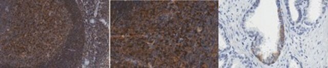
Evaluated by Immunohistochemistry in human tonsil and human bone marrow tissues.

Immunohistochemistry Analysis: A 1:250 dilution of this antibody detected PD-1 in human tonsil and human bone marrow tissues.

Other Notes

Concentration: Please refer to lot specific datasheet.

Disclaimer

Unless otherwise stated in our catalog or other company documentation accompanying the product(s), our products are intended for research use only and are not to be used for any other purpose, which includes but is not limited to, unauthorized commercial uses, in vitro diagnostic uses, ex vivo or in vivo therapeutic uses or any type of consumption or application to humans or animals.

Ähnliche Artikel vergleichen

Vollständigen Vergleich anzeigen

Unterschiede anzeigen

1 of 1

Dieser Artikel
SAB3500123MABC1115MABC1120
conjugate

unconjugated

conjugate

unconjugated

conjugate

unconjugated

conjugate

unconjugated

species reactivity

human

species reactivity

rat, human, mouse

species reactivity

human

species reactivity

human

biological source

mouse

biological source

mouse

biological source

mouse

biological source

mouse

clone

EH33.1F9.G4.5, monoclonal

clone

12A7D7, monoclonal

clone

5H1, monoclonal

clone

366C.12.9E5.E10.19.1F8, monoclonal

Gene Information

human ... PDCD1(5133)

Gene Information

human ... PDCD1(5133)

Gene Information

human ... CD274(29126)

Gene Information

human ... PDCD1LG2(80380)

antibody form

purified immunoglobulin

antibody form

purified immunoglobulin

antibody form

purified immunoglobulin

antibody form

purified immunoglobulin


Still not finding the right product?

Probieren Sie unser Produkt-Auswahlhilfe, um Ihre Auswahl einzugrenzen


Lagerklasse

12 - Non Combustible Liquids

wgk

WGK 1

flash_point_f

Not applicable

flash_point_c

Not applicable


Analysenzertifikate (COA)

Suchen Sie nach Analysenzertifikate (COA), indem Sie die Lot-/Chargennummer des Produkts eingeben. Lot- und Chargennummern sind auf dem Produktetikett hinter den Wörtern ‘Lot’ oder ‘Batch’ (Lot oder Charge) zu finden.

Besitzen Sie dieses Produkt bereits?

In der Dokumentenbibliothek finden Sie die Dokumentation zu den Produkten, die Sie kürzlich erworben haben.

Die Dokumentenbibliothek aufrufen


Questions

Reviews

No rating value

Active Filters