Skip to Content
Merck
All Photos(3)

Documents

MABC94

Sigma-Aldrich

Anti-Sphingosine 1-phosphate receptor 1 (S1P1) Antibody, clone 8B7.1

clone 8B7.1, from mouse

Synonym(s):

Sphingosine 1-phosphate receptor 1, S1P receptor 1, S1P1, Endothelial differentiation G-protein coupled receptor 1, Sphingosine 1-phosphate receptor Edg-1, S1P receptor Edg-1, CD363

Sign Into View Organizational & Contract Pricing


About This Item

UNSPSC Code:
12352203
eCl@ss:
32160702
NACRES:
NA.41

biological source

mouse

Quality Level

antibody form

purified immunoglobulin

antibody product type

primary antibodies

clone

8B7.1, monoclonal

species reactivity

mouse, human, rat

technique(s)

immunohistochemistry: suitable
western blot: suitable

isotype

IgG2a

NCBI accession no.

UniProt accession no.

shipped in

wet ice

target post-translational modification

unmodified

Gene Information

human ... S1PR1(1901)

General description

Sphingosine 1-phosphate receptor 1(S1PR1 or S1P1), otherwise known as EDG1, is a G-protein coupled receptor which is activated by lysosphingolipid sphingosine 1-phosphate (S1P) and lysophosphatidic acid (LPA). It is coupled to Gi-protein and MAP kinase signaling pathways, and may cooperate with other S1P receptors to regulate various cellular processes such as differentiation, actin remodeling, and migration. S1P1 is expressed in epithelial cells, fibroblasts, melanocytes, and smooth muscle cells.

Immunogen

GST-tagged recombinant protein corresponding to mouse Sphingosine 1-phosphate receptor 1 (S1P1).

Application

Research Category
Apoptosis & Cancer
Research Sub Category
GPCR, cAMP/cGMP & Calcium Signaling
This Anti-Sphingosine 1-phosphate receptor 1 (S1P1) Antibody, clone 8B7.1 is validated for use in Western Blotting, IHC for the detection of Sphingosine 1-phosphate receptor 1 (S1P1).
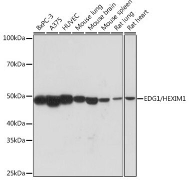
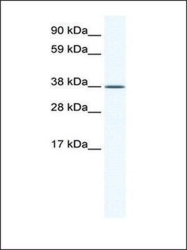
Western Blot Analysis: A 1:500 dilution from a representative lot detected Sphingosine 1-phosphate receptor 1 (S1P1) in human brain tissue lysate.

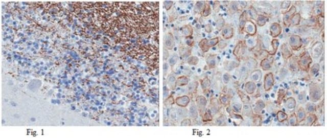
Immunohistochemistry Analysis: A 1:300 dilution from a representative lot detected Sphingosine 1-phosphate receptor 1 (S1P1) in rat choroid plexus tissue.

Quality

Evaluated by Western Blot in mouse brain tissue lysate.

Western Blot Analysis: A 1:1,000 diluiton of this antibody detected Sphingosine 1-phosphate receptor 1 (S1P1) in 10 µg of mouse brain tissue lysate.

Target description

~40 kDa observed

Physical form

Format: Purified
Protein G Purified
Purified mouse monoclonal IgG2a supernatant in buffer containing 0.1 M Tris-Glycine (pH 7.4), 150 mM NaCl with 0.05% sodium azide.

Storage and Stability

Stable for 1 year at 2-8°C from date of receipt.

Analysis Note

Control
Mouse brain tissue lysate

Disclaimer

Unless otherwise stated in our catalog or other company documentation accompanying the product(s), our products are intended for research use only and are not to be used for any other purpose, which includes but is not limited to, unauthorized commercial uses, in vitro diagnostic uses, ex vivo or in vivo therapeutic uses or any type of consumption or application to humans or animals.

Not finding the right product?  

Try our Product Selector Tool.

Storage Class Code

12 - Non Combustible Liquids

WGK

WGK 1

Flash Point(F)

Not applicable

Flash Point(C)

Not applicable


Certificates of Analysis (COA)

Search for Certificates of Analysis (COA) by entering the products Lot/Batch Number. Lot and Batch Numbers can be found on a product’s label following the words ‘Lot’ or ‘Batch’.

Already Own This Product?

Find documentation for the products that you have recently purchased in the Document Library.

Visit the Document Library

Alhaji H Janneh et al.
Cell reports, 41(10), 111742-111742 (2022-12-09)
Crosstalk between metabolic and signaling events that induce tumor metastasis remains elusive. Here, we determine how oncogenic sphingosine 1-phosphate (S1P) metabolism induces intracellular C3 complement activation to enhance migration/metastasis. We demonstrate that increased S1P metabolism activates C3 complement processing through
Gustavo H Oliveira-Paula et al.
Science advances, 10(11), eadg9278-eadg9278 (2024-03-13)
Canonical Wnt and sphingosine-1-phosphate (S1P) signaling pathways are highly conserved systems that contribute to normal vertebrate development, with key consequences for immune, nervous, and cardiovascular system function; despite these functional overlaps, little is known about Wnt/β-catenin-S1P cross-talk. In the vascular
Jeffrey Thompson et al.
Aging and disease (2024-06-25)
Endothelial dysfunction and blood-brain barrier (BBB) leakage have been suggested as a fundamental role in the development of cerebral small vessel disease (SVD) pathology. However, the molecular and cellular mechanisms that link cerebral hypoxic hypoperfusion and BBB disruption remain elusive.
Sandeep K Singh et al.
Glia, 70(4), 712-727 (2021-12-28)
Astrocytes, the most abundant glial cells in the mammalian brain, directly associate with and regulate neuronal processes and synapses and are important regulators of brain development. Yet little is known of the molecular mechanisms that control the establishment of astrocyte

Our team of scientists has experience in all areas of research including Life Science, Material Science, Chemical Synthesis, Chromatography, Analytical and many others.

Contact Technical Service