
A4325
Monoclonal Anti-α-Adaptin antibody produced in mouse
clone 100/2, ascites fluid
Synonym(s):
Anti-AP-2
Sign Into View Organizational & Contract Pricing
All Photos(1)
About This Item
Pricing and availability is not currently available.
Recommended Products


biological source
mouse
conjugate
unconjugated
antibody form
ascites fluid
antibody product type
primary antibodies
clone
100/2, monoclonal
contains
15 mM sodium azide
species reactivity
bovine, human, rat
technique(s)
western blot: 1:200 using a crude preparation of synaptic vesicles obtained from rat cerebral cortex
isotype
IgG2a
UniProt accession no.
shipped in
dry ice
storage temp.
−20°C
target post-translational modification
unmodified
Gene Information
human ... AP2A1(160)
rat ... Ap2a1(308578)
General description
Clathrin-coated vesicles facilitate the selective internalization of membrane receptors for lysosomal enzymes from the trans-Golgi network to a pre-lysosomal compartment. Besides clathrin, coated vesicle populations contain the adaptor complexes AP-1 and AP-2, also known as HA-I (HA1) adaptor and HA-II (HA2) adaptor or Assemble Protein 1 (AP1) and Assemble Protein 2 (AP2), respectively. These adaptor proteins mediate the interaction between clathrin and membrane components.
Monoclonal Anti-a-Adaptin (AP-2) (mouse IgG2a isotype) is derived from the 100/2 hybridoma produced by the fusion of mouse myeloma cells and splenocytes from an immunized mouse. Adaptor related protein complex 2 subunit alpha 1 (AP2A1) codes for αA adaptin of the AP-2 complex. It is usually seen in clathrin coated vesicles.
The antibody reacts with polypeptides of ~100 kDa in bovine liver, human heart fibroblasts, and Madin-Darby bovine kidney cultured cells (MDBK), but not with any components in the 110-115 kDa range from these sources or from rat pheochromocytoma cultured cells (PC12), neuroblastoma, or astrocytes. This suggests that the 110 and 115 kDa polypeptides may be specific variants that occur only in some cell types of brain. The antibody stains the intact α-adaptor, as well as the 37 and 40 kDa fragments, but not the 63-66 kDa group of α-fragments obtained by trypsin cleavage of the α-subunit molecules.
Immunogen
AP-2 adaptor polypeptides from bovine brain.
Application
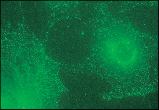
Immunofluorescence of MDCK cells fixed in methanol was performed using anti-AP1 at a dilution of 1:250 and also western blot at a dilution of 1:1000.
Monoclonal Anti-α-Adaptin antibody produced in mouse has been used in immunoblotting.
Mouse monoclonal anti-α-adaptin antibody has been used:
- as a positive control in immunoblotting assays
- as a primary antibody to detect AP2 levels in tetracycline-repressible stable cell lines
Biochem/physiol Actions
Adaptor related protein complex 2 subunit α 1 (AP2α1) is very essential for epidermal growth factor receptor (EGFR) nuclear translocation, induced by ultraviolet (UV).
Physical form
The product is provided as ascites fluid with 0.1% sodium azide as a preservative.
Disclaimer
Unless otherwise stated in our catalog or other company documentation accompanying the product(s), our products are intended for research use only and are not to be used for any other purpose, which includes but is not limited to, unauthorized commercial uses, in vitro diagnostic uses, ex vivo or in vivo therapeutic uses or any type of consumption or application to humans or animals.
Not finding the right product?
Try our Product Selector Tool.
Storage Class Code
10 - Combustible liquids
WGK
WGK 3
Flash Point(F)
Not applicable
Flash Point(C)
Not applicable
Choose from one of the most recent versions:
Already Own This Product?
Find documentation for the products that you have recently purchased in the Document Library.
Abstract 4049: AP2A1 is required for UV-induced EGFR nuclear translocation
Hsu SH
Cancer research, 4049-4049 (2011)
Juana M Del Valle et al.
The Journal of biological chemistry, 278(19), 17430-17437 (2003-03-07)
CD229 (Ly9) is a cell surface receptor selectively expressed on T and B lymphocytes, and it belongs to the CD150 receptor family. Like other receptors of this family, CD229 interacts with SAP/SH2D1a protein, mutation of which is responsible for the
Vincent Blot et al.
Retrovirology, 3, 62-62 (2006-09-19)
Retrovirus particles emerge from the assembly of two structural protein components, Gag that is translated as a soluble protein in the cytoplasm of the host cells, and Env, a type I transmembrane protein. Because both components are translated in different
Molecular cloning of novel transcripts of the adaptor-related protein complex 2 alpha 1 subunit (AP2A1) gene, using Next-Generation Sequencing
Adamopoulos PG, et al.
Gene, 678, 55-64 (2018)
R A Warren et al.
The Journal of biological chemistry, 273(27), 17056-17063 (1998-06-27)
Endocytosis of surface proteins through clathrin-coated pits requires an internalization signal in the cytoplasmic domain. Two types of internalization signal have been described: one requiring a tyrosine as the critical residue (tyrosine-based motif), and the other consisting of either two
Active Filters
Our team of scientists has experience in all areas of research including Life Science, Material Science, Chemical Synthesis, Chromatography, Analytical and many others.
Contact Technical Service