Skip to Content
Merck
All Photos(1)

Key Documents

904H-0

Sigma-Aldrich

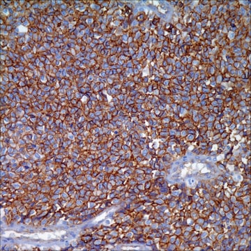
HMB-45 + MART-1 (Melan A) + Tyrosinase (HMB-45 + A103 + T311) Mouse Monoclonal Antibody

Sign Into View Organizational & Contract Pricing


About This Item

UNSPSC Code:
12352203
NACRES:
NA.41

biological source

mouse

Quality Level

100
500

conjugate

unconjugated

antibody form

culture supernatant

antibody product type

primary antibodies

clone

HMB-45+A103+T311, monoclonal

description

For In Vitro Diagnostic Use in Select Regions (See Chart)

form

buffered aqueous solution

species reactivity

human

packaging

bottle of 1.0 mL predilute (904H-07)
bottle of 7.0 mL predilute (904H-08)

manufacturer/tradename

Cell Marque

isotype

IgG1κ
IgG1
IgG2a

shipped in

wet ice

storage temp.

2-8°C

General description

Anti-HMB-45 identifies immature melanosomes. It shows greater specificity for malignant melanomas and metastatic melanomas. MART-1 (also known as Melan A) is a melanocyte differentiation antigen. It is present in melanocytes of normal skin and retina, nevi and in more than 85% of melanomas. Tyrosinase is an enzyme integral in the process of melanin synthesis, and found in 85% to 90% of malignant melanomas. Given these statistics, this cocktail is ideally suited to detection of melanomas and melanocytic lesions.

Quality


IVD

IVD

IVD

RUO

Physical form

Solution in Tris Buffer, pH 7.3-7.7, with 1% BSA and <0.1% Sodium Azide

Preparation Note

Download the IFU specific to your product lot and formatNote: This requires a keycode which can be found on your packaging or product label.

Other Notes

For Technical Service please contact: 800-665-7284 or email: service@cellmarque.com

Legal Information

Cell Marque is a trademark of Merck KGaA, Darmstadt, Germany

Not finding the right product?  

Try our Product Selector Tool.

Storage Class Code

12 - Non Combustible Liquids

WGK

WGK 1

Flash Point(F)

Not applicable

Flash Point(C)

Not applicable


Certificates of Analysis (COA)

Search for Certificates of Analysis (COA) by entering the products Lot/Batch Number. Lot and Batch Numbers can be found on a product’s label following the words ‘Lot’ or ‘Batch’.

Already Own This Product?

Find documentation for the products that you have recently purchased in the Document Library.

Visit the Document Library

Guy Orchard
British journal of biomedical science, 59(4), 196-202 (2003-02-08)
Incidence of malignant melanoma (MM) is rising rapidly throughout the Western world, and the number of melanocytic lesions removed for histological assessment has increased. MM can present with a myriad of histological appearances that make diagnosis problematic, particularly when dealing
Deepali Gupta et al.
The American journal of surgical pathology, 26(11), 1450-1457 (2002-11-01)
Malignant melanomas of the vagina are rare tumors. In this study we present the clinicopathologic features and immunohistochemical analysis of 26 such cases seen in our institution over a period of 30 years. The patients' age ranged from 38 to
L Vaggelli et al.
Tumori, 86(4), 346-348 (2000-10-04)
Elective lymph node dissection (ELND) for patients with malignant melanoma is still controversial. A possible alternative could be biopsy of the first tumor draining lymph node, the sentinel node (SN), which can be identified by means of radionuclide techniques. Our
T J de Vries et al.
The Journal of pathology, 193(1), 13-20 (2001-02-13)
With the recent availability of novel antibodies against melanoma antigens tyrosinase and MART-1, it is important to validate their usefulness in pathology practice and in screening patients for immunotherapy treatment. In the present study conducted by the Melanoma Cooperative Group
M L Prasad et al.
The American journal of surgical pathology, 25(6), 782-787 (2001-06-08)
Malignant melanomas of the oral and sinonasal mucosa are rare tumors. Amelanotic variants can, on occasion, be difficult to recognize by routine light microscopy. Immunohistochemical studies may be needed for a final diagnosis. A number of new monoclonal antibodies to

Articles

IHC antibodies enhance dermatopathology beyond H&E stained slides, improving techniques and applications for dermatological research.

IHC antibodies enhance dermatopathology beyond H&E stained slides, improving techniques and applications for dermatological research.

IHC antibodies enhance dermatopathology beyond H&E stained slides, improving techniques and applications for dermatological research.

IHC antibodies enhance dermatopathology beyond H&E stained slides, improving techniques and applications for dermatological research.

Our team of scientists has experience in all areas of research including Life Science, Material Science, Chemical Synthesis, Chromatography, Analytical and many others.

Contact Technical Service