Accéder au contenu
Merck
Toutes les photos(3)

Key Documents

MABS482

Sigma-Aldrich

Anti-PL Scramblase 1 Antibody, clone 1A8

clone 1A8, from mouse

Synonyme(s) :

Phospholipid scramblase 1, PL Scramblase 1, Ca(2+)-dependent phospholipid scramblase 1, Erythrocyte phospholipid scramblase, MmTRA1b

Se connecterpour consulter vos tarifs contractuels et ceux de votre entreprise/organisme


About This Item

Code UNSPSC :
12352203
eCl@ss :
32160702
Nomenclature NACRES :
NA.41

Source biologique

mouse

Niveau de qualité

Forme d'anticorps

purified antibody

Type de produit anticorps

primary antibodies

Clone

1A8, monoclonal

Espèces réactives

human, mouse

Technique(s)

immunohistochemistry: suitable
immunoprecipitation (IP): suitable
western blot: suitable

Isotype

IgG2bκ

Numéro d'accès NCBI

Numéro d'accès UniProt

Conditions d'expédition

wet ice

Modification post-traductionnelle de la cible

unmodified

Informations sur le gène

human ... PLSCR1(5359)

Description générale

Phospholipid scramblase 1 (UniProt O15162; also known as Ca(2+)-dependent phospholipid scramblase 1, Erythrocyte phospholipid scramblase, MmTRA1b, PL scramblase 1) is encoded by the PLSCR1 (also known as MMTRA1B) gene (Gene ID 5359) in human. Plasma membrane phospholipids are distributed asymmetrically between the inner and outer leaflets. Such asymmetrical distribution collapses in response to blood coagulation and apoptosis, resulting in phospholipid “scrambling” between the two leaflets of the plasma membrane. Flippases, floppases, and scramblases are three types enzymes known to mediate transbilayer lipid motion. Flippases and floppases function via ATP-dependent mechanism, while scramblases mediate transbilayer movement in a non-selective and energy-independent manner. Originally identified in 1996 as a 37 kDa erythrocyte type II transmembrane protein that mediates calcium-dependent membrane phospholipids redistribution, PL Scramblase 1 is the protein product encoded by the founding member of the PLSCR family of genes (PLSCR1-5). All PLSCR family members, with the exception of PLSCR2, possess a proline-rich N-terminal region containing PxxP and PPxY domains, a cysteine-rich region, a conserved calcium-binding domain (EF-hand-like), and a putative transmembrane region enriched in hydrophobic amino acids. In addition, PLSCR1 contains a nuclear localization signal (NLS) and a DNA-binding domain that are essential for its nuclear localization and associated nuclear function. In addition to PLSCRs, TMEM16 and XKR family members have also been reported to mediate scramblase activity.

Immunogène

Recombinant protein corresponding to human PL Scramblase 1.

Application

Anti-PL Scramblase 1 Antibody, clone 1A8 is an antibody against PL Scramblase 1 for use in Western Blotting, Immunohistochemistry, Immunoprecipitation.
Research Category
Signaling
Research Sub Category
Signaling Neuroscience
Western Blotting Analysis: 1.0 µg/mL from a representative lot detected PL Scramblase 1 in 10 µg of A549 cell lysate.
Immunohistochemistry Analysis: A 1:50 dilution from a representative lot detected PL Scramblase 1 in mouse colon tissue.
Western Blotting Analysis: A representative lot detected PL scramblase 1 in platelets and lung tissue from wild-type and PLSCR3-/-, but not PLSCR1-/-, mice, while little or no PL scramblase 1 was detected in adipocyte or muscle samples from wild-type or the knockout animals (Wiedmer, T., et al. (2004). Proc Natl Acad Sci USA. 101(36):13296-13301).
Western Blotting Analysis: A representative lot detected retroviral-mediated ectopic expression of murine PLSCR1 in SCF-ER-Hoxb8-immortalized myeloid progenitor cells from PLSCR1−/− mice (Chen, C.W., et al. (2011). J Leukoc Biol. 90(2):221-233).
Immunprecipitation Analysis: A representative lot immunoprecipitated PL scramblase 1 from murine bone marrow-derived mast cells (BMMCs) following IgE receptor activation. Tyrosine phosphorylation of PL scramblase 1 was then checked by Western blotting with clone 4G10 (Kassas, A., et al. (2014). PLoS One. 9(10):e109800).

Qualité

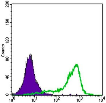
Evaluated by Western Blotting in HeLa cell lysate.

Western Blotting Analysis: 1.0 µg/mL of this antibody detected PL Scramblase 1 in 10 µg of HeLa cell lysate.

Description de la cible

~35 kDa observed. Uncharacterized band(s) may appear in some lysates.

Forme physique

Format: Purified
Protein G Purified
Purified mouse monoclonal IgG2bκ antibody in buffer containing 0.1 M Tris-Glycine (pH 7.4), 150 mM NaCl with 0.05% sodium azide.

Stockage et stabilité

Stable for 1 year at 2-8°C from date of receipt.

Autres remarques

Concentration: Please refer to lot specific datasheet.

Clause de non-responsabilité

Unless otherwise stated in our catalog or other company documentation accompanying the product(s), our products are intended for research use only and are not to be used for any other purpose, which includes but is not limited to, unauthorized commercial uses, in vitro diagnostic uses, ex vivo or in vivo therapeutic uses or any type of consumption or application to humans or animals.

Vous ne trouvez pas le bon produit ?  

Essayez notre Outil de sélection de produits.

Code de la classe de stockage

12 - Non Combustible Liquids

Classe de danger pour l'eau (WGK)

WGK 1

Point d'éclair (°F)

Not applicable

Point d'éclair (°C)

Not applicable


Certificats d'analyse (COA)

Recherchez un Certificats d'analyse (COA) en saisissant le numéro de lot du produit. Les numéros de lot figurent sur l'étiquette du produit après les mots "Lot" ou "Batch".

Déjà en possession de ce produit ?

Retrouvez la documentation relative aux produits que vous avez récemment achetés dans la Bibliothèque de documents.

Consulter la Bibliothèque de documents

Genomewide CRISPR knockout screen identified PLAC8 as an essential factor for SADS-CoVs infection.
Tse, et al.
Proceedings of the National Academy of Sciences of the USA, 119, e2118126119-e2118126119 (2023)

Notre équipe de scientifiques dispose d'une expérience dans tous les secteurs de la recherche, notamment en sciences de la vie, science des matériaux, synthèse chimique, chromatographie, analyse et dans de nombreux autres domaines..

Contacter notre Service technique