Accéder au contenu
MilliporeSigma
Toutes les photos(2)

Documents

PRS3459

Sigma-Aldrich

Anti-Caspase-1 (ab1) antibody produced in rabbit

affinity isolated antibody, buffered aqueous solution

Synonyme(s) :

Anti-Caspase 1, apoptosis-related cysteine peptidase (Interleukin 1, beta, convertase), Anti-ICE, Anti-IL-1β converting enzyme

Se connecterpour consulter vos tarifs contractuels et ceux de votre entreprise/organisme


About This Item

Numéro MDL:
Code UNSPSC :
12352203
Nomenclature NACRES :
NA.43

Source biologique

rabbit

Conjugué

unconjugated

Forme d'anticorps

affinity isolated antibody

Type de produit anticorps

primary antibodies

Clone

polyclonal

Forme

buffered aqueous solution

Espèces réactives

human

Technique(s)

immunofluorescence: suitable
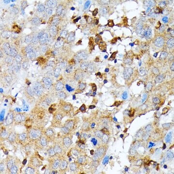
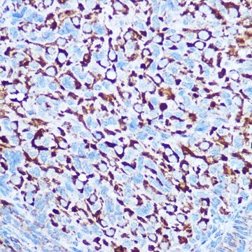
immunohistochemistry: suitable
indirect ELISA: suitable
western blot: suitable

Numéro d'accès UniProt

Conditions d'expédition

dry ice

Température de stockage

−20°C

Modification post-traductionnelle de la cible

unmodified

Informations sur le gène

human ... CASP1(834)

Description générale

Caspase-1 is also known as interleukin-1β converting enzyme (ICE). It is located on chromosome 11q22.2. It has 10 exons, an N terminal CARD (caspase activation and recruitment domain), a large P20 subunit and a small P10 subunit. It is present in leukocytes, monocytes and epithelial cells.

Immunogène

a 16 amino acid peptide from near the carboxy-terminus of human Caspase-1.

Application

Anti-Caspase-1 (ab1) antibody produced in rabbit has been used in western blotting and immunofluorescence staining.

Actions biochimiques/physiologiques

Caspase-1 can transform the pro-inflammatory cytokines, interleukin-1β (IL-1β) and IL-18 into their active forms. It participates in pyroptosis. It is overexpressed in the aorta of coronary atherosclerosis patients. It helps in host cell survival by inducing membrane biogenesis to restore the impairment caused by pore-forming toxins.

Liaison

The action of this antibody can be blocked using blocking peptide SBP3459.

Forme physique

Solution in phosphate buffered saline containing 0.02% sodium azide

Clause de non-responsabilité

Unless otherwise stated in our catalog or other company documentation accompanying the product(s), our products are intended for research use only and are not to be used for any other purpose, which includes but is not limited to, unauthorized commercial uses, in vitro diagnostic uses, ex vivo or in vivo therapeutic uses or any type of consumption or application to humans or animals.

Not finding the right product?  

Try our Outil de sélection de produits.

Produit(s) apparenté(s)

Réf. du produit
Description
Tarif

Code de la classe de stockage

10 - Combustible liquids

Classe de danger pour l'eau (WGK)

WGK 2

Point d'éclair (°F)

Not applicable

Point d'éclair (°C)

Not applicable


Certificats d'analyse (COA)

Recherchez un Certificats d'analyse (COA) en saisissant le numéro de lot du produit. Les numéros de lot figurent sur l'étiquette du produit après les mots "Lot" ou "Batch".

Déjà en possession de ce produit ?

Retrouvez la documentation relative aux produits que vous avez récemment achetés dans la Bibliothèque de documents.

Consulter la Bibliothèque de documents

Qi Chen et al.
The Journal of biological chemistry, 290(43), 26051-26058 (2015-09-13)
Autophagy is a cellular process that controls and executes the turnover of dysfunctional organelles and misfolded or abnormally aggregated proteins. Phosphatase and tensin homologue deleted on chromosome 10 (PTEN) activates the initiation of autophagy. Autophagosomes migrate along acetylated microtubules to
CASP1 (caspase 1, apoptosis-related cysteine peptidase (interleukin 1, beta, convertase))
Kumar Y, et al.
Atlas of Genetics and Cytogenetics in Oncology and Haematology (2007)
Pyroptosis, a novel mechanism implicated in cataracts
Jin X, et al.
Molecular Medicine Reports, 18(2), 2277-2285 (2018)
Monocyte Caspase-1 Is Released in a Stable, Active High Molecular Weight Complex Distinct from the Unstable Cell Lysate-Activated Caspase-1
Shamaa OR, et al.
PLoS ONE, 10(11) (2015)
Guibin Xu et al.
Aging, 8(5), 977-985 (2016-05-30)
Excessive deposition of extracellular matrix proteins in renal tissues causes renal fibrosis and renal function failure. Mammalian cells primarily use the autophagy-lysosome system to degrade misfolded/aggregated proteins and dysfunctional organelles. MAP1S is an autophagy activator and promotes the biogenesis and

Notre équipe de scientifiques dispose d'une expérience dans tous les secteurs de la recherche, notamment en sciences de la vie, science des matériaux, synthèse chimique, chromatographie, analyse et dans de nombreux autres domaines..

Contacter notre Service technique