Skip to Content
MilliporeSigma
All Photos(3)

Documents

MABD51

Sigma-Aldrich

Anti-Brn-2 (POU3F2) Antibody, clone 8C4.2

clone 8C4.2, from mouse

Synonym(s):

POU domain, class 3, transcription factor 2, Brain-specific homeobox/POU domain protein 2, Brain-2, Brn-2, Nervous system-specific octamer-binding transcription factor N-Oct-3, Octamer-binding protein 7, Oct-7, Octamer-binding transcription factor 7, OTF

Sign Into View Organizational & Contract Pricing


About This Item

UNSPSC Code:
12352203
eCl@ss:
32160702
NACRES:
NA.41

biological source

mouse

Quality Level

antibody form

purified immunoglobulin

antibody product type

primary antibodies

clone

8C4.2, monoclonal

species reactivity

rat, human

technique(s)

immunohistochemistry: suitable (paraffin)
western blot: suitable

isotype

IgG1κ

NCBI accession no.

UniProt accession no.

shipped in

wet ice

target post-translational modification

unmodified

Gene Information

human ... POU3F2(5454)

General description

Brn-2 (POU domain, class 3, transcription factor 2; Oct-7; N-Oct-3; or POU3F2) is a member of a large family of POU-domain transcription factors. The highly homologous POU domain contains a POU-specific domain at the N-terminus, a POU-homeo domain at the C-terminus and an interconnecting linker region. The POU domain binds to the DNA sequence 5’-ATGCAAAT-3’. Brn-2 is highly expressed in the developing CNS and may play a role in neurogenesis, particularly in the development of the hypothalamus. Previous studies have also suggested that Brn-2 integrates signals downstream of the BRAF/MAP kinase and Wnt/β-catenin pathways, and Brn-2 may be involved in the transformation and proliferation of melanomas.

Immunogen

GST-tagged recombinant protein corresponding human Brn-2 (POU3F2).

Application

Anti-Brn-2 (POU3F2) Antibody, clone 8C4.2 detects level of Brn-2 (POU3F2) & has been published & validated for use in Western Blotting, IHC(P).
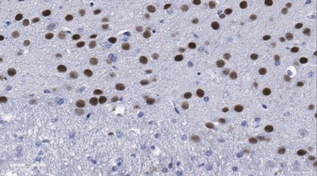
Immunohistochemistry Analysis: A 1:50 dilution from a representative lot detected Brn-2 in normal rat brain tissue, in Purkinje, basket, and glial cells of rat cerebellum tissue, in neurons of rat cerebellum tissue, in neurons and glial cells of rat frontal cortex tissue, and in neurons of human pons tissue.

Quality

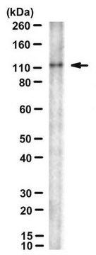
Evaluated by Western Blot in SK-N-MC cell lysates.

Western Blot Analysis: A 1:2,000 dilution of this antibody detected Brn-2 (POU3F2) in 10 µg of SK-N-MC cell lysates.

Target description

~54 kDa observed.
The calculated molecular weight is 47 kDa Brn-2 (POU3F2) may be observed at ~50-55 kDa in some cell lysates (Wolfe, A., et al., (2002). Molecular Endocrinology. 16(3):435-449).

Physical form

Format: Purified

Analysis Note

Control
SK-N-MC cell lysates

Not finding the right product?  

Try our Product Selector Tool.

Storage Class Code

12 - Non Combustible Liquids

WGK

WGK 1

Flash Point(F)

Not applicable

Flash Point(C)

Not applicable


Certificates of Analysis (COA)

Search for Certificates of Analysis (COA) by entering the products Lot/Batch Number. Lot and Batch Numbers can be found on a product’s label following the words ‘Lot’ or ‘Batch’.

Already Own This Product?

Find documentation for the products that you have recently purchased in the Document Library.

Visit the Document Library

Meitetsu Masawa et al.
The American journal of pathology, 192(6), 847-861 (2022-04-04)
Although recent reports have revealed the importance of the inactivation of both RB1 and TP53 in the transformation from lung adenocarcinoma into neuroendocrine carcinoma (NEC), the requirements for complete transformation into NEC have not been elucidated. To investigate alterations in
Zhongyuan Bao et al.
Oxidative medicine and cellular longevity, 2021, 6338722-6338722 (2021-12-03)
Traumatic brain injury (TBI) causes a high rate of mortality and disability, and its treatment is still limited. Loss of neurons in damaged area is hardly rescued by relative molecular therapies. Based on its disease characteristics, we transplanted human embryonic
Yilin Feng et al.
STAR protocols, 4(3), 102346-102346 (2023-07-08)
In glioma modeling, existing organoid protocols lack the ability to replicate glioma cell invasion and interaction with normal brain tissue. Here, we present a protocol for generating in vitro brain disease models using human-induced pluripotent- or embryonic-stem-cell-derived cerebral organoids (COs). We
Waseem K Raja et al.
PloS one, 17(12), e0277532-e0277532 (2022-12-02)
There are currently no preventive or disease-modifying therapies for Parkinson's Disease (PD). Failures in clinical trials necessitate a re-evaluation of existing pre-clinical models in order to adopt systems that better recapitulate underlying disease mechanisms and better predict clinical outcomes. In
Aaron Gordon et al.
Nature neuroscience, 24(3), 331-342 (2021-02-24)
Human stem-cell-derived models provide the promise of accelerating our understanding of brain disorders, but not knowing whether they possess the ability to mature beyond mid- to late-fetal stages potentially limits their utility. We leveraged a directed differentiation protocol to comprehensively

Our team of scientists has experience in all areas of research including Life Science, Material Science, Chemical Synthesis, Chromatography, Analytical and many others.

Contact Technical Service