ECM506
QCM Chemotaxis Cell Migration Assay, 24-well (5 µm), colorimetric
This 3 um QCM Chemotaxis Assay 24-well plate -colorimetric is performed in a Migration Chamber, based on the Boyden chamber principle.
Synonym(s):
Cell migration assay, Chemotaxis assay
Sign Into View Organizational & Contract Pricing
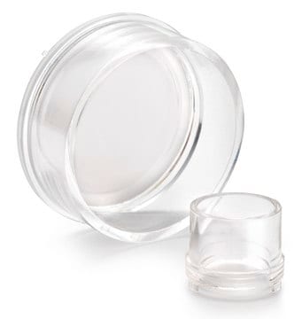
All Photos(1)
About This Item
UNSPSC Code:
12352207
eCl@ss:
32161000
NACRES:
NA.84
Recommended Products
Quality Level
species reactivity (predicted by homology)
all
manufacturer/tradename
Chemicon®
QCM
technique(s)
activity assay: suitable
cell based assay: suitable
detection method
colorimetric
shipped in
wet ice
Related Categories
General description
Also available: Cell Comb Scratch Assay! Get biochemical data from a scratch assay!Click Here
Introduction
Cell migration is a fundamental function of normal cellular processes, including embryonic development, angiogenesis, wound healing, immune response, and inflammation (1, 2). Most cells are sized from 30 to 50 µm can migrate through 3 to 10 µm pore. Microporous membrane inserts are widely used for cell migration and invasion assays. The most widely accepted of which is the Boyden Chamber assay. The Boyden Chamber system uses a hollow plastic chamber, sealed at one end with a porous membrane. This chamber is suspended over a larger well which may contain medium and/or chemoattractants. Cells are placed inside the Chamber and allowed to migrate through the pores, to the other side of the membrane. Migratory cells are then stained and counted. Most migration assays utilize an 8 µm pore size, as this is appropriate for most cell types, e.g. epithelial and fibroblast cells. The Millipore 5 µm QCM Chemotaxis Assay 24-well- colorimetric utilizes a 5 µm pore size, which is appropriate for a subset of fibroblast cells or cancer cells such as NIH-3T3 and MDA-MAB 231 cells. Cells migrated toward chemoattractants were measured by crystal violet, a nucleic dye, and quantified by spectrophotometers. The system may be adapted to study different types of cell migration, including haptotaxis, random migration, and chemokinesis.
The Millipore QCM 5 µm Chemotaxis Assay 24-well-colorimetric provides a quick and efficient system for quantitative determination of various factors on cell migration, including screening of pharmacological agents, evaluation of integrins or other adhesion receptors responsible for cell migration, or analysis of gene function in transfected cells.
In addition, Chemicon continues to provide numerous migration, invasion, and adhesion products including:
• QCM 8µm 24-well Chemotaxis Cell Migration Assays (ECM508, 509)
• QCM 5µm 24-well Chemotaxis Cell Migration Assay- Fluorometric (ECM507)
• QCM 8µm 96-well Chemotaxis Cell Migration Assay (ECM510)
• QCM 5µm 96-well Chemotaxis Cell Migration Assay (ECM512)
• QCM 3µm 96-well Chemotaxis Cell Migration Assay (ECM515)
• QCM 96-well ECMatrix Cell Invasion Assay (ECM555)
• QCM 96-well Collagen-based Cell Invasion Assay (ECM556)
Application
The Millipore 5 µm QCM Chemotaxis Assay 24-well-colorimetric is performed in a Migration Chamber, based on the Boyden chamber principle. The 5 µm pore size of this assay′s Boyden chambers is appropriate for studying a subset of fibroblast or cancer cell migration. The quantitative nature of this assay is useful for screening of pharmacological agents. Each kit provides sufficient materials for the evaluation of 24 samples.
The Millipore 5 µm QCM Chemotaxis Assay 24-well-colorimetric is intended for research use only; not for diagnostic applications.
The Millipore 5 µm QCM Chemotaxis Assay 24-well-colorimetric is intended for research use only; not for diagnostic applications.
Packaging
24 wells
Components
Sterile 5 µm 24-well Cell Migration Plate Assembly: (Part No. 2005708) Two 24-well plates with 12 inserts per plate (24 inserts total/kit).
Cell Stain: (Part No. 90144) one 20 ml.
Extraction buffer: (Part No. 90145) one 20ml.
Cotton Swaps: (Part No. 10202) 50 each.
Forceps: (Part No. 10203) One each.
Cell Stain: (Part No. 90144) one 20 ml.
Extraction buffer: (Part No. 90145) one 20ml.
Cotton Swaps: (Part No. 10202) 50 each.
Forceps: (Part No. 10203) One each.
Storage and Stability
The 5 µm 24-well Cell Migration Plate and forceps are stored at room temperature. Cell stain and extraction buffer can be store at 2° to 8°C up to the expiration date. Do not freeze any components.
Legal Information
Accutase is a registered trademark of Innovative Cell Technologies, Inc.
CHEMICON is a registered trademark of Merck KGaA, Darmstadt, Germany
Disclaimer
Unless otherwise stated in our catalog or other company documentation accompanying the product(s), our products are intended for research use only and are not to be used for any other purpose, which includes but is not limited to, unauthorized commercial uses, in vitro diagnostic uses, ex vivo or in vivo therapeutic uses or any type of consumption or application to humans or animals.
Signal Word
Danger
Hazard Statements
Precautionary Statements
Hazard Classifications
Eye Irrit. 2 - Flam. Liq. 2
Storage Class Code
3 - Flammable liquids
Flash Point(F)
53.6 °F
Flash Point(C)
12 °C
Certificates of Analysis (COA)
Search for Certificates of Analysis (COA) by entering the products Lot/Batch Number. Lot and Batch Numbers can be found on a product’s label following the words ‘Lot’ or ‘Batch’.
Already Own This Product?
Find documentation for the products that you have recently purchased in the Document Library.
Customers Also Viewed
Dola Das et al.
Fibrogenesis & tissue repair, 7, 9-9 (2014-06-12)
C5a and its cognate receptor, C5a receptor (C5aR), key elements of complement, are critical modulators of liver immunity and fibrosis. However, the molecular mechanism for the cross talk between complement and liver fibrosis is not well understood. C5a is a
Arash Aghajani Nargesi et al.
American journal of physiology. Renal physiology, 317(5), F1142-F1153 (2019-08-29)
Scattered tubular-like cells (STCs) contribute to repair neighboring injured renal tubular cells. Mitochondria mediate STC biology and function but might be injured by the ambient milieu. We hypothesized that the microenviroment induced by the ischemic and metabolic components of renovascular
Tatsuya Hasegawa et al.
Scientific reports, 11(1), 2613-2613 (2021-01-30)
Apical periodontitis (AP) is an acute or chronic inflammatory disease caused by complex interactions between infected root canal and host immune system. It results in the induction of inflammatory mediators such as chemokines and cytokines leading to periapical tissue destruction.
Christopher M Ferguson et al.
Cells, 10(4) (2021-04-04)
Percutaneous transluminal renal angioplasty (PTRA) confers clinical and mortality benefits in select 'high-risk' patients with renovascular disease (RVD). Intra-renal-delivered extracellular vesicles (EVs) released from mesenchymal stem/stromal cells (MSCs) protect the kidney in experimental RVD, but have not been compared side-by-side
Alfonso Eirin et al.
Stem cell research, 47, 101877-101877 (2020-06-28)
Mesenchymal stromal/stem cell (MSC)-derived extracellular vesicles (EVs) shuttle select MSC contents and are endowed with an ability to repair ischemic tissues. We hypothesized that exposure to cardiovascular risk factors may alter the microRNA cargo of MSC-derived EVs, blunting their capacity
Our team of scientists has experience in all areas of research including Life Science, Material Science, Chemical Synthesis, Chromatography, Analytical and many others.
Contact Technical Service


