Skip to Content
Merck
All Photos(2)

Documents

L2020

Sigma-Aldrich

Mouse Laminin

from Engelbreth-Holm-Swarm murine sarcoma basement membrane, 0.2 μm filtered, liquid, 1-2 mg/mL, suitable for cell culture

Synonym(s):

Laminin

Sign Into View Organizational & Contract Pricing


About This Item

CAS Number:
MDL number:
UNSPSC Code:
12352202
NACRES:
NA.75

product name

Laminin from Engelbreth-Holm-Swarm murine sarcoma basement membrane, 1-2 mg/mL in Tris-buffered saline, 0.2 μm filtered, BioReagent, suitable for cell culture

biological source

mouse (Engelbreth-Holm-Swarm mouse sarcoma basement membrane)

Quality Level

product line

BioReagent

form

aqueous solution

mol wt

A subunit 400 kDa
B1 subunit 210 kDa
B2 subunit 200 kDa

packaging

pkg of 1 mg

concentration

1-2 mg/mL in Tris-buffered saline

technique(s)

cell culture | mammalian: suitable

surface coverage

1‑2 μg/cm2

impurities

Microbial Contamination, passes test

NCBI accession no.

UniProt accession no.

Binding Specificity

Peptide Source: Collagen

shipped in

dry ice

storage temp.

−20°C

Looking for similar products? Visit Product Comparison Guide

General description

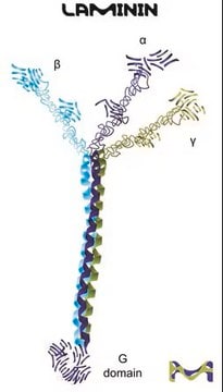
Laminins are non-collagenous adhesive glycoproteins with a substantial molecular weight. They are heterotrimeric proteins with three disulfide-linked polypeptides known as the α, β, and γ chains. The multiple polypeptide domains of laminins can bind to multiple cell surface receptors, including several integrin dimers, dystroglycan and syndecans, and other ECM components. This extracellular matrix multidomain trimeric glycoprotein is the main component of basal lamina that supports adhesion, proliferation and differentiation.

Application

Laminin from Engelbreth-Holm-Swarm murine sarcoma basement membrane has been used in:
  • insulin-producing cell (IPC) differentiation.
  • Infection inhibition assays.
  • Proliferation Assay.
Laminin supports growth and differentiation of many cell types including epithelial, endothelial, neural, muscle and liver cells. It is recommended for use as a cell culture substratum at 1-2 μg/cm2. The optimal concentration does depend on cell type as well as the application or research objectives.

Biochem/physiol Actions

Laminin proteins are integral components of structural scaffolding in animal tissues. They associate with type IV collagen via entactin and perlecan and bind to cell membranes through integrin receptors, dystroglycan glycoprotein complexes and Lutheran blood group glycoproteins. Laminin has active domains for collagen binding, cell adhesion, heparin binding, and neurite outgrowth fragment.

Components

Laminin is an extracellular matrix multidomain trimeric glycoprotein, and is the main non-collagenous component of basal lamina that supports adhesion, proliferation and differentiation. Laminin is composed of both A, B1 and B2 chains, which are connected by many disulfide bonds. This laminin product was isolated from mouse Engelbreth-Holm-Swarm tumor.

Caution

It is recommended to store this product in working aliquots at -20°C.

Preparation Note

This product is supplied at a concentration of 1 − 2 mg/mL in 50 mM Tris-HCl, with 150 mM NaCl. It is filtered using a 0.2μm filter. Thaw this solution slowly before use at 2-8°C to avoid gel formation. For use as a coating, dilute in a balanced salt solution, coat culture surface with a minimal volume and incubate at 37°C for two hours. Wash 3 times with PBS before plating cells. Laminin coatings can be stored for one month at 2-8°C.

related product

Product No.
Description
Pricing

Storage Class Code

11 - Combustible Solids

WGK

WGK 3

Flash Point(F)

Not applicable

Flash Point(C)

Not applicable

Personal Protective Equipment

dust mask type N95 (US), Eyeshields, Gloves

Certificates of Analysis (COA)

Search for Certificates of Analysis (COA) by entering the products Lot/Batch Number. Lot and Batch Numbers can be found on a product’s label following the words ‘Lot’ or ‘Batch’.

Already Own This Product?

Find documentation for the products that you have recently purchased in the Document Library.

Visit the Document Library

Axonal pathfinding: Extracellular matrix role
Letourneau, P
Encyclopedia of Neuroscience, 1, 1139-1145 (2009)
Serotype-specific entry of dengue virus into liver cells: identification of the 37-kilodalton/67-kilodalton high-affinity laminin receptor as a dengue virus serotype 1 receptor.
Thepparit C and Smith DR.
Journal of Virology, 78(22), 12647-12656 (2004)
Agnese Loda et al.
Nature communications, 8(1), 690-690 (2017-09-28)
Xist is indispensable for X chromosome inactivation. However, how Xist RNA directs chromosome-wide silencing and why some regions are more efficiently silenced than others remains unknown. Here, we explore the function of Xist by inducing ectopic Xist expression from multiple
Tobias Bergström et al.
Biochimica et biophysica acta, 1840(8), 2526-2532 (2014-01-28)
It is becoming increasingly apparent that the extracellular matrix acts as an important regulator of the neural stem niche. Previously we found that neural stem and progenitor cells (NSPCs) derived from the early postnatal subventricular zone of mice adhere to
Fibronectin and laminin promote differentiation of human mesenchymal stem cells into insulin producing cells through activating Akt and ERK.
Lin HY, et al.
Journal of Biomedical Science, 17:56 (2010)

Articles

The extracellular matrix (ECM) and its attachment factor components are discussed in this article in relation to their function in structural biology and their availability for in vitro applications.

The extracellular matrix (ECM) and its attachment factor components are discussed in this article in relation to their function in structural biology and their availability for in vitro applications.

The extracellular matrix (ECM) and its attachment factor components are discussed in this article in relation to their function in structural biology and their availability for in vitro applications.

The extracellular matrix (ECM) and its attachment factor components are discussed in this article in relation to their function in structural biology and their availability for in vitro applications.

See All

Protocols

Coating surfaces with laminin for culturing cells requires specific conditions for optimal results. Protocols for coating coverslips to culture neurospheres and general cell culture are included.

Coating surfaces with laminin for culturing cells requires specific conditions for optimal results. Protocols for coating coverslips to culture neurospheres and general cell culture are included.

Coating surfaces with laminin for culturing cells requires specific conditions for optimal results. Protocols for coating coverslips to culture neurospheres and general cell culture are included.

Coating surfaces with laminin for culturing cells requires specific conditions for optimal results. Protocols for coating coverslips to culture neurospheres and general cell culture are included.

Our team of scientists has experience in all areas of research including Life Science, Material Science, Chemical Synthesis, Chromatography, Analytical and many others.

Contact Technical Service