Skip to Content
Merck
All Photos(1)

Documents

A5968

Sigma-Aldrich

Anti-AP-1 antibody produced in rabbit

affinity isolated antibody, buffered aqueous solution

Synonym(s):

Ap-1 Antibody, Ap1 Antibody, Ap1 Antibody - Anti-AP-1 antibody produced in rabbit, Anti-c-Jun

Sign Into View Organizational & Contract Pricing


About This Item

MDL number:
UNSPSC Code:
12352203
NACRES:
NA.41

biological source

rabbit

conjugate

unconjugated

antibody form

affinity isolated antibody

antibody product type

primary antibodies

clone

polyclonal

form

buffered aqueous solution

mol wt

antigen 39 kDa

species reactivity

human

technique(s)

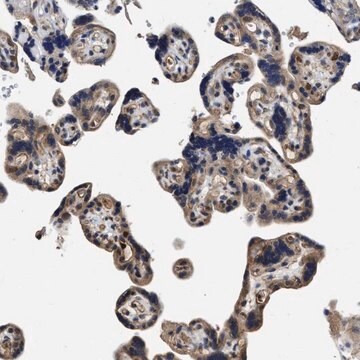
immunohistochemistry (formalin-fixed, paraffin-embedded sections): 1:200 using formalin-fixed, paraffin-embedded sections of human colon carcinoma
microarray: suitable
western blot: 1:200 using HeLa nuclear extract

UniProt accession no.

shipped in

dry ice

storage temp.

−20°C

target post-translational modification

unmodified

Gene Information

human ... JUN(3725)

General description

Activation Protein transcription factor 1 (AP-1) is a leucine zipper transcription factor that recognizes a specific palindromic sequence present in several promoters and enhancers. AP-1 can be induced by several growth and apoptosis-related signaling pathways. Jun proteins are also involved in metastatic, inflammatory and stress responses.
Anti-AP-1/c-Jun recognizes an epitope located on the c-Jun DNA binding domain. This epitope is highly conserved in c-Jun, Jun B and Jun D proteins of chicken, mouse, rat, and human. By immunoblotting, the antibody reacts specifically with AP-1/c-Jun (a single band or occasionally a doublet at 39kDa region). Additional bands of lower molecular weight may be observed. Staining of AP-1/c-Jun band(s) is inhibited by the AP-1/c-Jun immunizing peptide (amino acid residues 246-263).
Activation Protein transcription factor 1 (AP-1) is a regulatory leucine zipper transcription factor. AP-1 is composed of either a homodimer of two Jun protein molecules or a heterodimer of one Jun and one Fos (Proto-oncogenes) molecules.

Immunogen

synthetic peptide corresponding to amino acids 246-263 of human c-Jun with N-terminally added lysine, conjugated to KLH.

Application

Anti-AP-1 antibody has been used in
  • immunohistochemistry
  • immunochemistry
  • western blotting

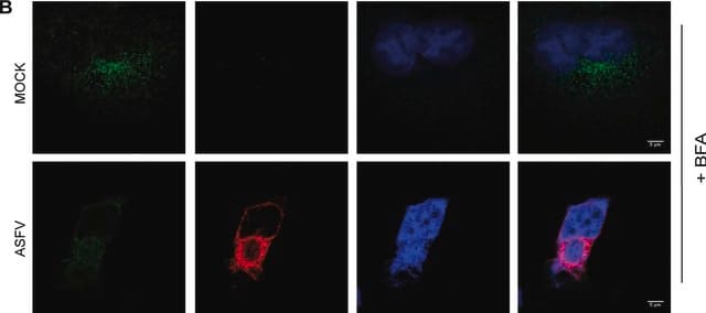
Immunofluorescence of Hela cells was performed using monoclonal anti-AP1 (clone 100/3) as the primary antibody.

Biochem/physiol Actions

Activation Protein transcription factor 1 (AP-1) recognizes a specific sequence (TPA response element, a palindromic TRE sequence TGA(C/G) TCA or related sequences) present in many promoter and enhancer gene regions. Regulation of AP-1 is achieved by changes in the expression of Jun and Fos proteins in the dimer, by phosphorylation at both Ser63 and Ser73 by c-Jun N-terminal kinases (JNK) and by interaction with a variety of transcriptional coactivators like C-Jun activation domain-binding protein-1 (JAB1). AP-1 is induced by a variety of signals including those eliciting proliferation, differentiation and apoptosis.

Physical form

Solution in 0.01 M phosphate buffered saline, pH 7.4, containing 1% bovine serum albumin and 15 mM sodium azide.

Disclaimer

Unless otherwise stated in our catalog or other company documentation accompanying the product(s), our products are intended for research use only and are not to be used for any other purpose, which includes but is not limited to, unauthorized commercial uses, in vitro diagnostic uses, ex vivo or in vivo therapeutic uses or any type of consumption or application to humans or animals.

Not finding the right product?  

Try our Product Selector Tool.

Storage Class Code

10 - Combustible liquids

WGK

WGK 3

Flash Point(F)

Not applicable

Flash Point(C)

Not applicable


Certificates of Analysis (COA)

Search for Certificates of Analysis (COA) by entering the products Lot/Batch Number. Lot and Batch Numbers can be found on a product’s label following the words ‘Lot’ or ‘Batch’.

Already Own This Product?

Find documentation for the products that you have recently purchased in the Document Library.

Visit the Document Library

Yongshun Tan et al.
Cell biology international, 45(8), 1654-1665 (2021-03-25)
TET1 mediates demethylation in tumors, but its role in diabetic nephropathy (DN), a prevalent diabetic complication, is unclear. We attempted to probe the possible mechanism of TET1 in DN. A DN rat model was established and verified by marker detection and
Swathy Krishna et al.
Frontiers in physiology, 14, 1152576-1152576 (2023-05-14)
Duchenne muscular dystrophy (DMD) is a progressive muscle disease that results in muscle wasting, wheelchair dependence, and eventual death due to cardiac and respiratory complications. In addition to muscle fragility, dystrophin deficiency also results in multiple secondary dysfunctions, which may
AP-1 as a regulator of cell life and death
Shaulian E and Karin M
Nature Cell Biology, 4(5), E131-E131 (2002)
Adaptor and Clathrin Exchange at the Plasma Membrane and trans-Golgi Network.
Xufeng, W.
Molecular and Cellular Biology, 14, 516-528 (2002)
Ui Joung Youn et al.
Biological & pharmaceutical bulletin, 34(6), 890-893 (2011-06-02)
Tumor necrosis factor α (TNF-α), which is a primary cytokine responsible for inflammatory responses in skin, induces the synthesis of matrix metalloproteinase-9 (MMP-9), which causes skin aging. The protective effects of 3-deoxysappanchalcone against TNF-α-induced damage was investigated using human skin

Our team of scientists has experience in all areas of research including Life Science, Material Science, Chemical Synthesis, Chromatography, Analytical and many others.

Contact Technical Service