Skip to Content
Merck
All Photos(4)

Key Documents

17-10032

Sigma-Aldrich

ChIPAb+ Trimethyl-Histone H3 (Lys36) - ChIP Validated Antibody and Primer Set, rabbit monoclonal

from rabbit

Synonym(s):

H3K36me3, Histone H3 (tri methyl K36), H3 histone family, member T, histone 3, H3, histone cluster 3, H3

Sign Into View Organizational & Contract Pricing


About This Item

UNSPSC Code:
12352203
eCl@ss:
32160702
NACRES:
NA.32
clone:
monoclonal
application:
ChIP
DB
IP
WB
cell based assay
species reactivity:
human, chicken
technique(s):
ChIP: suitable
cell based assay: suitable
dot blot: suitable
immunoprecipitation (IP): suitable
western blot: suitable
citations:
10

biological source

rabbit

Quality Level

antibody form

purified immunoglobulin

clone

monoclonal

species reactivity

human, chicken

manufacturer/tradename

ChIPAb+
Upstate®

technique(s)

ChIP: suitable
cell based assay: suitable
dot blot: suitable
immunoprecipitation (IP): suitable
western blot: suitable

isotype

IgG

NCBI accession no.

UniProt accession no.

shipped in

dry ice

General description

All ChIPAb+ antibodies are individually validated for chromatin precipitation, every lot, every time. Each ChIPAb+ antibody set includes control primers (tested every lot by qPCR) to biologically validate your IP results in a locus-specific context. The qPCR protocol and primer sequences are provided, allowing researchers to validate ChIP protocols when using our antibody in their chromatin context. Each set also includes a negative control antibody to ensure specificity of the ChIP reaction.
The ChIPAb+ Trimethyl-Histone H3 (Lys36) set includes the Trimethyl-Histone H3 (Lys36) antibody, a negative control antibody (normal rabbit IgG), and qPCR primers which amplify a 147 bp region of human BDNF intron. The Trimethyl-Histone H3 (Lys36) and negative control antibodies are supplied in a scalable "per ChIP" reaction size and can be used to functionally validate the precipitation of Trimethyl-Histone H3 (Lys36)-associated chromatin.
Histones are highly conserved proteins that serve as the structural scaffold for the organization of nuclear DNA into chromatin. The four core histones, H2A, H2B, H3, and H4, assemble into an octamer (2 molecules of each). Subsequently, 146 base pairs of DNA are wrapped around the octamer, forming a nucleosome, the basic subunit of chromatin. Histones are modified post-translationally by the actions of enzymes in both the nucleus and cytoplasm. These modifications regulate DNA transcription, repair, recombination, and replication. The most commonly studied modifications are acetylation, phosphorylation, methylation, and ubiquitination. These modifications can alter local chromatin architecture, or recruit trans-acting factors that recognize specific histone modifications (the "histone code" hypothesis). The modifications occur predominantly on the N-terminal and C-terminal tails that extend beyond the nucleosome core particle. Methylation of histone H3 on Lys36 (H3K36me2/3) is tightly associated with actively transcribed genes, and this modification is found primarily within the coding region, suggesting H3K36 methylation is necessary for efficient RNA polymerase II elongation and processivity.

Specificity

Recognizes Trimethyl-Histone H3 (Lys36), Mr ~17 kDa.
Wide reactivity with other species is expected.

Immunogen

Epitope: Trimethylated Lys36
KLH-conjugated, synthetic peptide containing the sequence ....GVme3KKP…, in which me3K corresponds to human trimethyl-histone H3 (Lys36).

Application

Chromatin Immunoprecipitation:
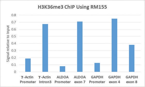
Sonicated chromatin prepared from HeLa cells (1 X 10⁶ cell equivalents per IP) were subjected to chromatin immunoprecipitation using 2 µg of either Normal rabbit IgG or 2 µL Anti-trimethyl-Histone H3 (Lys36) and the Magna ChIP A Kit (Cat. # 17-610). Successful immunoprecipitation of trimethyl-Histone H3 (Lys36) associated DNA fragments was verified by qPCR using ChIP Primers, BDNF Intron as a positive locus, and GAPDH promoter primers as a negative locus (Please see figures). Data is presented as percent input of each IP sample relative to input chromatin for each amplicon and ChIP sample as indicated.
Please refer to the EZ-Magna ChIP A (Cat. # 17-408) or EZ-ChIP (Cat. # 17-371) protocol for experimental details.

Western Blot Analysis and Peptide Inhibition:
Representative lot data.
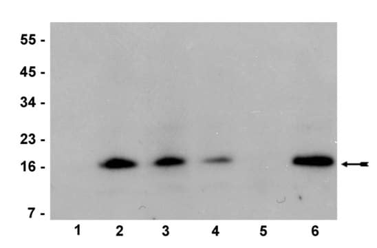
Recombinant Histone H3 (Catalog # 14-411, lane 1) and chicken Core Histones (Catalog # 13-107, lane 2) were resolved by electrophoresis, transferred to nitrocellulose and probed with antitrimethyl-Histone H3 (Lys36) (1:1000 dilution) or anti-trimethyl-Histone H3 (Lys36) pre-adsorbed with 1mM histone H3 peptides containing the following modifications:
Lane 3: monomethyl-lysine 36
Lane 4: dimethyl-lysine 36
Lane 5: trimethyl-lysine 36
Lane 6: unmodified
Proteins were visualized using a goat anti-rabbit secondary antibody conjugated to HRP and a chemiluminescence detection system. (Please see figures).

Peptide Dot Blot Analysis:
Representative lot data.
A dilution series of Histone H3 peptides containing the following modifications was made:
Column 1: unmodified
Column 2: monomethyl-lysine 36
Column 3: dimethyl-lysine 36
Column 4: trimethyl-lysine 36
2 μL of each dilution was spotted onto a PVDF membrane and probed with antitrimethyl-Histone H3 (Lys36), (1:1000 dilution).
Peptides were visualized using a goat anti-rabbit secondary conjugated to HRP and a chemiluminescence detection system (Please see figures).

Research Category
Epigenetics & Nuclear Function
Research Sub Category
Histones
This ChIPAb+ Trimethyl-Histone H3 (Lys36) -ChIP Validated Antibody & Primer Set conveniently includes the antibody & the specific control PCR primers.

Packaging

25 assays per set. Recommended use: ~2 μL of antibody per chromatin immunoprecipitation (dependent upon biological context).

Quality

Chromatin Immunoprecipitation:
Sonicated chromatin prepared from HeLa cells (1 X 10⁶ cell equivalents per IP) were subjected to chromatin immuno-precipitation using 2 µg of either Normal Rabbit IgG or 2 µL Anti-trimethyl-Histone H3 (Lys36) and the Magna ChIP® A Kit (Cat. # 17-610).
Successful immunoprecipitation of trimethyl-Histone H3 (Lys36) associated DNA fragments was verified by qPCR using ChIP Primers, BDNF Intron (Please see figures).
Please refer to the EZ-Magna ChIP A (Cat. # 17-408) or EZ-ChIP (Cat. # 17-371) protocol for experimental details.

Target description

~17 kDa

Physical form

Anti-Trimethyl-Histone H3 (Lys36) (rabbit monoclonal IgG). One vial containing 50 μL of protein A purified IgG in a solution containing 0.07 M Tris-glycine, 0.105 M NaCl, pH 7.4, 0.035% sodium azide and 30% glycerol. Store at -20°C.

Normal Rabbit IgG. One vial containing 125 µg Rabbit IgG in 125 µL of storage buffer containing 0.05% sodium azide. Store at -20°C.

ChIP Primers, BDNF Intron. One vial containing 75 μL of 5 μM of each primer specific for human BDNF intron. Store at -20°C.
FOR: ACCCCAACCTCTAACAGCATTA
REV: TGTCTCTCAGCAGTCTTGCATT
Format: Purified
Protein A purified

Storage and Stability

Stable for 1 year at -20°C from date of receipt. Handling Recommendations: Upon first thaw, and prior to removing the cap, centrifuge the vial and gently mix the solution. Aliquot into microcentrifuge tubes and store at -20°C. Avoid repeated freeze/thaw cycles, which may damage IgG and affect product performance. Note: Variability in freezer temperatures below -20°C may cause glycerol containing solutions to become frozen during storage.

Analysis Note

Control
Includes negative control rabbit IgG antibody and primers specific for human BDNF Intron.

Legal Information

MAGNA CHIP is a registered trademark of Merck KGaA, Darmstadt, Germany
UPSTATE is a registered trademark of Merck KGaA, Darmstadt, Germany

Disclaimer

Unless otherwise stated in our catalog or other company documentation accompanying the product(s), our products are intended for research use only and are not to be used for any other purpose, which includes but is not limited to, unauthorized commercial uses, in vitro diagnostic uses, ex vivo or in vivo therapeutic uses or any type of consumption or application to humans or animals.

Storage Class Code

10 - Combustible liquids


Certificates of Analysis (COA)

Search for Certificates of Analysis (COA) by entering the products Lot/Batch Number. Lot and Batch Numbers can be found on a product’s label following the words ‘Lot’ or ‘Batch’.

Already Own This Product?

Find documentation for the products that you have recently purchased in the Document Library.

Visit the Document Library

Jing Yang et al.
Evidence-based complementary and alternative medicine : eCAM, 2019, 6479136-6479136 (2019-07-06)
The incidence of cardiac dysfunction after myocardial infarction (MI) continues to increase despite advances in treatment. Excessive myocardial fibrosis plays a vital role in the development of adverse cardiac remodeling and deterioration of cardiac function. Understanding the molecular and cellular
Keren Martínez-Aguilar et al.
Frontiers in plant science, 7, 653-653 (2016-06-01)
To survive in adverse conditions, plants have evolved complex mechanisms that "prime" their defense system to respond and adapt to stresses. Their competence to respond to such stresses fundamentally depends on its capacity to modulate the transcriptome rapidly and specifically.
Hazem E Hassan et al.
Epigenetics, 11(10), 740-749 (2016-09-03)
Curcumin and its analogs exhibited antileukemic activity either as single agent or in combination therapy. Dimethoxycurcumin (DMC) is a more metabolically stable curcumin analog that was shown to induce the expression of promoter-methylated genes without reversing DNA methylation. Accordingly, co-treatment
Pei Han et al.
Nature, 514(7520), 102-106 (2014-08-15)
The role of long noncoding RNA (lncRNA) in adult hearts is unknown; also unclear is how lncRNA modulates nucleosome remodelling. An estimated 70% of mouse genes undergo antisense transcription, including myosin heavy chain 7 (Myh7), which encodes molecular motor proteins
Anurag Kulkarni et al.
Cell chemical biology, 24(11), 1377-1387 (2017-10-03)
The human retrovirus HTLV-1 causes a hematological malignancy or neuroinflammatory disease in ∼10% of infected individuals. HTLV-1 primarily infects CD4+ T lymphocytes and persists as a provirus integrated in their genome. HTLV-1 appears transcriptionally latent in freshly isolated cells; however

Our team of scientists has experience in all areas of research including Life Science, Material Science, Chemical Synthesis, Chromatography, Analytical and many others.

Contact Technical Service