Skip to Content
Merck
All Photos(2)

Documents

SAB5500182

Sigma-Aldrich

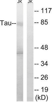
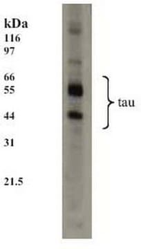
Anti-TAU antibody, Rabbit monoclonal

recombinant, expressed in proprietary host, clone SP70, affinity isolated antibody

Synonym(s):

Anti-DDPAC, Anti-FTDP-17, Anti-MAPTL, Anti-MSTD, Anti-MTBT1, Anti-MTBT2, Anti-PPND, Anti-PPP1R103, Anti-TAU, Anti-tau-40

Sign Into View Organizational & Contract Pricing


About This Item

UNSPSC Code:
12352203
NACRES:
NA.41

biological source

rabbit

Quality Level

recombinant

expressed in proprietary host

conjugate

unconjugated

antibody form

affinity isolated antibody

antibody product type

primary antibodies

clone

SP70, monoclonal

species reactivity

human (tested)

technique(s)

flow cytometry: 1:100
immunohistochemistry: 1:100

isotype

IgG

UniProt accession no.

shipped in

wet ice

storage temp.

2-8°C

target post-translational modification

unmodified

Gene Information

human ... MAPT(4137)

General description

Tau is a neuronal microtubule associated protein found predominantly on axons. The function of rau is to promote tubulin polymerization and stabilize microtubules, but it also serves to link certain signaling pathways to the cytoskeleton. Tau, in its hyperphosphorylated form, is the major component of paired helical filaments (PHF) and neurofibrillary lesions in Alzheimer′s disease (AD) brain. Hyperphosphorylation impairs the microtubule binding function of tau, resulting in the destabilization of microtubules in AD brains, ultimately leading to the degeneration of the affected neurons. Hyperphosphorylated tau is also found in a range of other central nervous system disorders. Numerous serine/threonine kinases, including GSK3 beta, PKA, Cdk5, and casein kinase II can phosphorylate tau. Alternate splicing of tau mRNA and differential phosphorylation contributes to the heterogeneity of Tau.

Immunogen

Synthetic peptide corresponding to N-terminus of human tau protein.

Features and Benefits

Evaluate our antibodies with complete peace of mind. If the antibody does not perform in your application, we will issue a full credit or replacement antibody. Learn more.

Physical form

0.1 ml rabbit monoclonal antibody purified by protein A/G in PBS/1% BSA buffer pH 7.6 with less than 0.1% sodium azide.

Disclaimer

Unless otherwise stated in our catalog or other company documentation accompanying the product(s), our products are intended for research use only and are not to be used for any other purpose, which includes but is not limited to, unauthorized commercial uses, in vitro diagnostic uses, ex vivo or in vivo therapeutic uses or any type of consumption or application to humans or animals.

Not finding the right product?  

Try our Product Selector Tool.

Storage Class Code

10 - Combustible liquids

WGK

WGK 2

Flash Point(F)

Not applicable

Flash Point(C)

Not applicable


Certificates of Analysis (COA)

Search for Certificates of Analysis (COA) by entering the products Lot/Batch Number. Lot and Batch Numbers can be found on a product’s label following the words ‘Lot’ or ‘Batch’.

Already Own This Product?

Find documentation for the products that you have recently purchased in the Document Library.

Visit the Document Library

Siwei Chu et al.
npj aging, 8(1), 16-16 (2022-12-17)
Intestinal epithelial cells are critical for gastrointestinal homeostasis. However, their function declines during aging. The aging-related loss of organ performance is largely driven by the increase in senescent cells. To date, the hallmarks and molecular mechanisms related to cellular senescence
Yuancheng Li et al.
ACS biomaterials science & engineering, 5(7), 3595-3605 (2019-07-08)
Alzheimer's disease (AD) is a growing global healthcare burden affecting the aging population and society. Given the lack of effective treatment to AD, early detection at the prodromal stage and timely monitoring of changes during progression are considered the best

Our team of scientists has experience in all areas of research including Life Science, Material Science, Chemical Synthesis, Chromatography, Analytical and many others.

Contact Technical Service