Skip to Content
Merck
All Photos(6)

Documents

06-536

Sigma-Aldrich

Anti-Bak Antibody, NT

Upstate®, from rabbit

Synonym(s):

Apoptosis regulator BAK, BCL2-antagonist/killer 1, BCL2-like 7 protein, Bcl-2 homologous antagonist/killer, Bcl-2-like 7 protein, pro-apoptotic protein BAK

Sign Into View Organizational & Contract Pricing


About This Item

UNSPSC Code:
12352203
eCl@ss:
32160702
NACRES:
NA.41

biological source

rabbit

Quality Level

antibody form

purified antibody

antibody product type

primary antibodies

clone

polyclonal

species reactivity

mouse, rat, human

manufacturer/tradename

Upstate®

technique(s)

immunocytochemistry: suitable
immunohistochemistry: suitable
immunoprecipitation (IP): suitable
western blot: suitable

isotype

IgG

NCBI accession no.

UniProt accession no.

shipped in

dry ice

target post-translational modification

unmodified

Gene Information

human ... BAK1(578)

General description

Bak or Bcl2 homologous antagonist is a member of the Bcl2 family of proteins. The Bcl-2 related proteins interact with one another through the formation of homo and heterodimers. The susceptibility of cells to apoptotic stimuli is thought to be controlled by the relative ratios of the different Bcl2 family proteins. Bak has been demonstrated to accelerate the rate of apoptosis in growth factor deprived murine lymphoid, neuronal and fibroblastic cell lines. This protein localizes to mitochondria, and functions to induce apoptosis. It interacts with and accelerates the opening of the mitochondrial voltage-dependent anion channel, which leads to a loss in membrane potential and the release of cytochrome c. Bak also interacts with the tumor suppressor P53 after exposure to cell stress.

Specificity

Specific for Bak, MW ~28 kDa.

Immunogen

Epitope: N-terminus
Synthetic peptide (SEEQVAQQDTEEVFRSC) corresponding to amino acid residues 23-38 of human Bak with a cysteine residue added to the C-terminus for conjugation to KLH.

Application

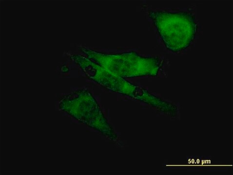
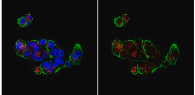
Detect Bak using this Anti-Bak Antibody, NT validated for use in IC, IH, IP & WB.
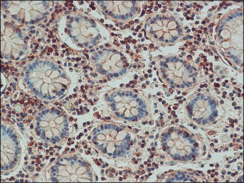
Immunohistochemistry (Paraffin):
5 μg/mL of a previous lot detected Bak in paraffin-embedded rat kidney tissue.

Immunoprecipitation:
4 μg of a previous lot immunoprecipitated Bak from 500 μg mouse 3T3 cell lysate.
Research Category
Apoptosis & Cancer
Research Sub Category
BCL2 & Inhibition

Quality

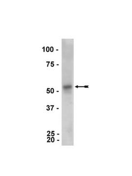
Evaluated by Western Blot on HEK293 lysates.

Western Blotting Analysis:
1:500 dilution of this antibody detected Bak on 10 µg of HEK293 lysates.

Target description

28 kDa

Linkage

Replaces: 04-433

Physical form

Format: Purified
Protein A purified
Purified rabbit polyclonal in storage buffer containing 0.1 M Tris-glycine, pH 7.4, 0.15 M NaCl, 0.05% sodium azide. Frozen liquid.

Storage and Stability

Stable for 1 year at -20ºC from date of receipt.

Analysis Note

Control
Positive Antigen Control: Catalog #12-301, non-stimulated A431 cell lysate. Add 2.5µL of 2-mercaptoethanol/100µL of lysate and boil for 5 minutes to reduce the preparation. Load 20µg of reduced lysate per lane for minigels.

Other Notes

Concentration: Please refer to the Certificate of Analysis for the lot-specific concentration.

Legal Information

UPSTATE is a registered trademark of Merck KGaA, Darmstadt, Germany

Disclaimer

Unless otherwise stated in our catalog or other company documentation accompanying the product(s), our products are intended for research use only and are not to be used for any other purpose, which includes but is not limited to, unauthorized commercial uses, in vitro diagnostic uses, ex vivo or in vivo therapeutic uses or any type of consumption or application to humans or animals.

Not finding the right product?  

Try our Product Selector Tool.

Storage Class Code

12 - Non Combustible Liquids

WGK

WGK 1

Flash Point(F)

Not applicable

Flash Point(C)

Not applicable


Certificates of Analysis (COA)

Search for Certificates of Analysis (COA) by entering the products Lot/Batch Number. Lot and Batch Numbers can be found on a product’s label following the words ‘Lot’ or ‘Batch’.

Already Own This Product?

Find documentation for the products that you have recently purchased in the Document Library.

Visit the Document Library

Molecular regulation of DNA damage-induced apoptosis in neurons of cerebral cortex.
Martin, LJ; Liu, Z; Pipino, J; Chestnut, B; Landek, MA
Cerebral Cortex (1991)
Anna V Miller et al.
PloS one, 8(4), e60685-e60685 (2013-04-12)
Paclitaxel (Taxol)-induced cell death requires the intrinsic cell death pathway, but the specific participants and the precise mechanisms are poorly understood. Previous studies indicate that a BH3-only protein BIM (BCL-2 Interacting Mediator of cell death) plays a role in paclitaxel-induced
Nuria López-Royuela et al.
Biochemical pharmacology, 79(12), 1746-1758 (2010-03-02)
Bcl-2 family proteins are key regulators of the intrinsic apoptotic pathway, either facilitating (Bax, Bak, BH3-only) or inhibiting (Bcl-2, Bcl-x(L), Mcl-1, A1) mitochondrial release of apoptogenic factors. The role of caspases in this process is a matter of controversy. We
Bim is the key mediator of glucocorticoid-induced apoptosis and of its potentiation by rapamycin in human myeloma cells.
Lopez-Royuela N, Balsas P, Galan-Malo P, Anel A, Marzo I, Naval J
Biochimica et Biophysica Acta null
Stephanie Einsele-Scholz et al.
Journal of cell science, 129(11), 2213-2223 (2016-04-15)
The pro-apoptotic multidomain Bcl-2 proteins Bax and Bak (also known as BAK1) are considered the gatekeepers of the intrinsic pathway of apoptosis by triggering the mitochondrial release of cytochrome c The role of the third Bax- and Bak-homologous multidomain protein

Our team of scientists has experience in all areas of research including Life Science, Material Science, Chemical Synthesis, Chromatography, Analytical and many others.

Contact Technical Service