Skip to Content
Merck
All Photos(3)

Key Documents

05-1459

Sigma-Aldrich

Anti-SMAD1 Antibody, clone AS22

clone AS22, from mouse

Synonym(s):

SMAD 1, SMAD family member 1, SMAD, mothers against DPP homolog 1, MAD, mothers against decapentaplegic homolog 1, Mad-related protein 1, Mothers against DPP homolog 1, Sma- and Mad-related protein 1, TGF-beta signaling protein 1, Transforming growth fac

Sign Into View Organizational & Contract Pricing


About This Item

UNSPSC Code:
12352203
eCl@ss:
32160702
NACRES:
NA.41

biological source

mouse

Quality Level

antibody form

purified antibody

antibody product type

primary antibodies

clone

AS22, monoclonal

species reactivity

mouse, human

technique(s)

immunofluorescence: suitable
western blot: suitable

isotype

IgG1κ

NCBI accession no.

UniProt accession no.

shipped in

wet ice

target post-translational modification

unmodified

Gene Information

human ... SMAD1(4086)

General description

Smad proteins are regulators of transcription which transduce signals from TGFβ Receptors. Smad proteins homotrimerize, and when activated, two distinct homotrimers assemble into a heterosextamer. Smad proteins fall into three classes. The receptor-regulated Smad proteins, Smad 1, 2, 3, 5, and 9 couple to specific receptors and are phosphorylated by those receptors. Phosphorylated receptor-regulated Smad proteins then bind to a co-Smad, such as Smad4/DPC4, and the complex moves to the nucleus where it associates with FAST-1 to stimulate target gene transcription. A third class of Smad proteins is the inhibitory group of Smad 6, 7, 8. Smad proteins have Mad-homology domains 1 and 2 (MH1 and MH2). MH1 domains are involved in DNA binding, while MH2 domains function in homotrimerization, receptor interaction and phosphorylation. The phosphorylation of Smad proteins is the regulatory signal in their activation, and can be monitored by the use of phosphorylation state-specific antibodies.

Specificity

Detects SMAD1
Other species were not tested.

Immunogen

GST-fusion protein corresponding to amino acids 1-110 of human SMAD1.

Application

Anti-SMAD1 Antibody, clone AS22 detects level of SMAD1 & has been published & validated for use in WB & IF.
Research Category
Signaling
Western Blot Analysis:
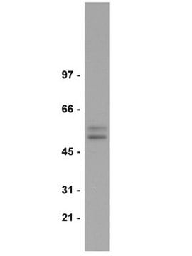
This lot detected SMAD1 at 1:500 dilution in HeLa cell lysate resolved via SDS-PAGE and transferred to PVDF.

Quality

Routinely evaluated by Western Blot

Western Blot Analysis:
This lot detected SMAD1 at 1:500 dilution in HeLa cell lysate resolved via SDS-PAGE and transferred to PVDF.

Target description

52 kDa

Linkage

Replaces: 04-1100

Physical form

Format: Purified
Protein G Purified
Purified mouse monoclonal in 0.1M Tris-Glycine (pH7.4), 150mM NaCl with 0.05% NaN3.

Storage and Stability

Stable for 1 year at 2-8°C from date of receipt.
Handling Recommendations: Upon receipt, and prior to removing the cap, centrifuge the vial and gently mix the solution.

Other Notes

Concentration: Please refer to the Certificate of Analysis for the lot-specific concentration.

Disclaimer

Unless otherwise stated in our catalog or other company documentation accompanying the product(s), our products are intended for research use only and are not to be used for any other purpose, which includes but is not limited to, unauthorized commercial uses, in vitro diagnostic uses, ex vivo or in vivo therapeutic uses or any type of consumption or application to humans or animals.

Not finding the right product?  

Try our Product Selector Tool.

Storage Class Code

12 - Non Combustible Liquids

WGK

WGK 1

Flash Point(F)

Not applicable

Flash Point(C)

Not applicable


Certificates of Analysis (COA)

Search for Certificates of Analysis (COA) by entering the products Lot/Batch Number. Lot and Batch Numbers can be found on a product’s label following the words ‘Lot’ or ‘Batch’.

Already Own This Product?

Find documentation for the products that you have recently purchased in the Document Library.

Visit the Document Library

Smita Amarnath et al.
Journal of cell science, 130(1), 119-131 (2016-04-02)
Many organs form by invaginating and rolling flat epithelial cell sheets into tubes. Invagination of the ventral midline of the neural plate forms the median hinge point (MHP), an event that elevates the neural folds and is essential for neural
Ariadne Cristiane Cabral Cruz et al.
Journal of applied oral science : revista FOB, 20(6), 628-635 (2013-01-19)
Bone morphogenetic protein type 2 (BMP-2) is a potent local factor, which promotes bone formation and has been used as an osteogenic supplement for mesenchymal stem cells. This study evaluated the effect of a recombinant BMP-2 as well as the
Ariadne Cristiane Cabral Cruz et al.
Journal of applied oral science : revista FOB, 27, e20180317-e20180317 (2019-02-28)
Bone morphogenetic protein type 2 (BMP-2) and retinoic acid (RA) are osteoinductive factors that stimulate endogenous mechanisms of bone repair which can be applied on management of osseous defects in oral and maxillofacial fields. Considering the different results of RA
Gregory J Heller et al.
Molecular therapy : the journal of the American Society of Gene Therapy, 29(5), 1883-1902 (2021-01-29)
Neonatal AAV9-gene therapy of the lysosomal enzyme galactosylceramidase (GALC) significantly ameliorates central and peripheral neuropathology, prolongs survival, and largely normalizes motor deficits in Twitcher mice. Despite these therapeutic milestones, new observations identified the presence of multiple small focal demyelinating areas in
Yi Sui et al.
British journal of cancer, 125(7), 1003-1015 (2021-07-12)
Metastasis is the major cause of treatment failure and cancer-related deaths in prostate cancer (PCa) patients. Our previous study demonstrated that a CD44+ subpopulation isolated from PCa cells or tumours possesses both stem cell properties and metastatic potential, serving as

Our team of scientists has experience in all areas of research including Life Science, Material Science, Chemical Synthesis, Chromatography, Analytical and many others.

Contact Technical Service