Skip to Content
Merck
All Photos(1)

Key Documents

345860

Sigma-Aldrich

Anti-Glial Fibrillary Acidic Protein Rat mAb (2.2B10)

liquid, clone 2.2B10, Calbiochem®

Synonym(s):

Anti-GFAP

Sign Into View Organizational & Contract Pricing


About This Item

UNSPSC Code:
12352203
NACRES:
NA.41

biological source

rat

Quality Level

antibody form

purified antibody

antibody product type

primary antibodies

clone

2.2B10, monoclonal

form

liquid

contains

≤0.1% sodium azide as preservative

species reactivity

bovine, rat, mouse, human

manufacturer/tradename

Calbiochem®

storage condition

OK to freeze
avoid repeated freeze/thaw cycles

isotype

IgG2a

shipped in

wet ice

storage temp.

−20°C

target post-translational modification

unmodified

Gene Information

human ... GCM1(8521)

General description

Purified rat monoclonal antibody. Recognizes the ~55 kDa glial fibrillary acidic protein (GFAP).
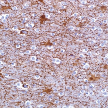
Recognizes GFAP in astrocytes induced by a variety of CNS injuries in human and rat brain tissues.
This Anti-Glial Fibrillary Acidic Protein Rat mAb (2.2B10) is validated for use in ELISA, Immunoblotting, Frozen Sections, IP, Paraffin Sections for the detection of Glial Fibrillary Acidic Protein.

Immunogen

Bovine Brain
bovine GFAP

Application

ELISA (0.1-0.5 µg/ml)

Immunoblotting (0.1-0.5 µg/ml)

Frozen Sections (10-50 µg/ml)

Immunoprecipitation (2-5 µg/ml)

Paraffin Sections (10-50 µg/ml, see comments)

Packaging

Please refer to vial label for lot-specific concentration.

Warning

Toxicity: Standard Handling (A)

Physical form

In PBS.

Reconstitution

Following initial thaw, aliquot and freeze (-20°C).

Analysis Note

Positive Control
Human or rat brain tissues

Other Notes

Product is not to be used for animal treatment, in vivo research, or in any other contact procedure with livestock. Stains reactive rodent and human brain astrocytes induced by a variety of central nervous system injuries. This antibody is suitable for immunohistochemical staining of Bouin′s-fixed, frozen, or paraffin-embedded tissue sections. Variables associated with assay conditions will dictate the proper working dilution.
Tohyama, T., et al. 1993. Am. J. Pathol.142, 871.
Lee, V.M., et al. 1984. J. Neurochem. 42, 25.

Legal Information

CALBIOCHEM is a registered trademark of Merck KGaA, Darmstadt, Germany

Not finding the right product?  

Try our Product Selector Tool.

Storage Class Code

11 - Combustible Solids

WGK

WGK 1

Flash Point(F)

Not applicable

Flash Point(C)

Not applicable


Certificates of Analysis (COA)

Search for Certificates of Analysis (COA) by entering the products Lot/Batch Number. Lot and Batch Numbers can be found on a product’s label following the words ‘Lot’ or ‘Batch’.

Already Own This Product?

Find documentation for the products that you have recently purchased in the Document Library.

Visit the Document Library

Michael S Petrik et al.
Neuromolecular medicine, 9(1), 83-100 (2006-11-23)
Gulf War illness (GWI) affects a significant percentage of veterans of the 1991 conflict, but its origin remains unknown. Associated with some cases of GWI are increased incidences of amyotrophic lateral sclerosis and other neurological disorders. Whereas many environmental factors
Dorin Sade Yazdi et al.
Proceedings of the National Academy of Sciences of the United States of America, 118(24) (2021-06-09)
High levels of homocysteine are reported as a risk factor for Alzheimer's disease (AD). Correspondingly, inborn hyperhomocysteinemia is associated with an increased predisposition to the development of dementia in later stages of life. Yet, the mechanistic link between homocysteine accumulation
Flora Guillot et al.
Journal of neuroinflammation, 12, 130-130 (2015-07-05)
Astrocytes, the most abundant cell population in mammal central nervous system (CNS), contribute to a variety of functions including homeostasis, metabolism, synapse formation, and myelin maintenance. White matter (WM) reactive astrocytes are important players in amplifying autoimmune demyelination and may
Tadasuke Tominaga et al.
PloS one, 14(3), e0213673-e0213673 (2019-03-12)
Primary and secondary traumatic brain injury (TBI) can cause tissue damage by inducing cell death pathways including apoptosis, necroptosis, and autophagy. However, similar pathways can also lead to senescence. Senescent cells secrete senescence-associated secretory phenotype proteins following persistent DNA damage
Marco Foddis et al.
Journal of cerebral blood flow and metabolism : official journal of the International Society of Cerebral Blood Flow and Metabolism, 40(2), 276-287 (2019-09-25)
Brain collateral circulation is an essential compensatory mechanism in response to acute brain ischemia. To study the temporal evolution of brain macro and microcollateral recruitment and their reciprocal interactions in response to different ischemic conditions, we applied a combination of

Our team of scientists has experience in all areas of research including Life Science, Material Science, Chemical Synthesis, Chromatography, Analytical and many others.

Contact Technical Service