rat, hamster, monkey, mouse, bovine, human, canine
concentration
~1 mg/mL
technique(s)
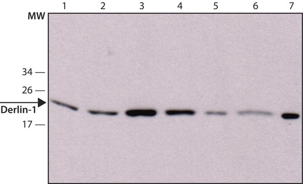
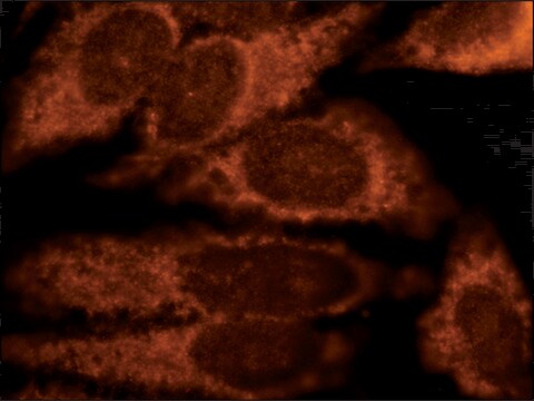
indirect immunofluorescence: 2.5-5 μg/mL using rat NRK cells western blot (chemiluminescent): 0.2-0.4 μg/mL using whole extract of human HeLa and mouse 3T3 cells.
Derlin-1 is a 22kDa hydrophobic protein that spans the lipid bilayer of the ER four times with its amino- and carboxy-terminus in the cytosol. It is expressed with high levels in liver, spleen, pancreas, lung, thymus, and ovary.
Derlin-1 shares human homology with yeast Der1p.
Immunogen
a synthetic peptide corresponding to the C-terminal region of human Derlin-1 with N-terminal added cysteine, conjugated to KLH. The corresponding sequence is identical in mouse.
Application
Anti-Derlin-1 antibody produced in rabbit has been used in:
immunostaining
co-immunoprecipitation
immunofluorescence
Biochem/physiol Actions
Derlin-1 can interact with peptide:N-glycanase (PNGase), a deglycosylating enzyme, bringing it close to misfolding dislocating glycoproteins.
Derlin-1 is required for the dislocation of misfolded proteins from the ER lumen to the cytosol, where they are destroyed by the ubiquitin-proteasome system. It interacts with PNGase, a deglycosylating enzyme, bringing it close to misfolding dislocating glycoproteins. It forms a membrane protein complex with VIMP ( (VCP-interacting membrane protein) and this complex serves as a receptor for p97. p97 interacts with several ubiquitin ligases, thus recruiting them to Derlin-1.
Physical form
Solution in 0.01 M phosphate buffered saline, pH 7.4, containing 15 mM sodium azide.
Disclaimer
Unless otherwise stated in our catalog or other company documentation accompanying the product(s), our products are intended for research use only and are not to be used for any other purpose, which includes but is not limited to, unauthorized commercial uses, in vitro diagnostic uses, ex vivo or in vivo therapeutic uses or any type of consumption or application to humans or animals.
The surveillance of the structural fidelity of the proteome is of utmost importance to all cells. The endoplasmic reticulum (ER) is the organelle responsible for proper folding and delivery of proteins to the secretory pathway. It contains a sophisticated protein
Molecular characterization and expression of DERL1 in bovine ovarian follicles and corpora lutea
Ndiaye K, et al.
Reproductive Biology and Endocrinology, 8(1), 94-94 (2010)
The functions of liver fatty acid binding protein 1 (FABP1) in the regulation of nonalcoholic fatty liver disease (NAFLD) have been previously established. However, how FABP1 expression is dynamically regulated in metabolic disorders is unclear. Previous studies have reported that
Proceedings of the National Academy of Sciences of the United States of America, 102(40), 14132-14138 (2005-09-28)
Misfolded proteins are eliminated from the endoplasmic reticulum (ER) by retrotranslocation into the cytosol, a pathway hijacked by certain viruses to destroy MHC class I heavy chains. The translocation of polypeptides across the ER membrane requires their polyubiquitination and subsequent
Various pathologies result from disruptions to or stress of endoplasmic reticulum (ER) homeostasis, such as Parkinson's disease and most neurodegenerative illnesses, diabetes, pulmonary fibrosis, viral infections, and cancers. A critical process in maintaining ER homeostasis is the selection of misfolded
Our team of scientists has experience in all areas of research including Life Science, Material Science, Chemical Synthesis, Chromatography, Analytical and many others.