Skip to Content
Merck
All Photos(1)

Key Documents

MAB3560-C

Sigma-Aldrich

Anti-8-Oxoguanine, clone 483.15 Antibody (Ascites Free)

clone 483.15, from mouse

Synonym(s):

8-Oxoguanine

Sign Into View Organizational & Contract Pricing


About This Item

UNSPSC Code:
12352203
eCl@ss:
32160702
NACRES:
NA.41

biological source

mouse

Quality Level

antibody form

purified immunoglobulin

antibody product type

primary antibodies

clone

483.15, monoclonal

species reactivity

rat, human, monkey, bovine, mouse

species reactivity (predicted by homology)

all

technique(s)

ELISA: suitable
immunocytochemistry: suitable
immunohistochemistry: suitable

isotype

IgMκ

shipped in

dry ice

target post-translational modification

unmodified

Gene Information

human ... OGG1(4968)

General description

8-Oxoguanine is a mutagenic oxidative damage product of guanine. Oxidatively damaged base 8-oxoguanine is generated in the DNA of all living organisms due to the presence of reactive oxygen species in cells. DNA damage threatens the health of the genetic information stored in every DNA molecule; however enzymes exist in cells to protect against the mutagenic effect of this lesion. 8-oxoguanine is one of the most abundant and well-characterized DNA lesions generated by oxidative stress. It has been estimated that ~180 guanines are oxidized to 8-oxoG per mammalian cell per day. 8-oxoG is a miscoding lesion that can cause G:C to T:A or T:A to G:C transversion mutations. This lesion accumulates in DNA with age and it has been loosly linked to several cancers and diseases, such as Alzheimer′s and Parkinson′s.

Specificity

8-oxoguanine. By competitive ELISA the monoclonal showed no reactivity with dGMP, dAMP, dCMP or TMP in micromolar concentrations. Competition was only observed when the concentrations were increased to the millimolar range.
Target DNA modification is not species-specific.

Immunogen

8-oxoguanine adsorbed on to alumina.

Application

Immunohistochemistry Analysis: A representative lot detected cells with 8-oxoguanine (8-oxoG) immunoreactivity in the central veins (CV) and portal triads (PT) regions of paraffin-embedded kettle liver sections (García-Vaquero, M., et al. (2012). Animal.6(9):1435-1443).
Immunohistochemistry Analysis: A representative lot detected increased nuclear 8-oxoguanine (8-oxoG) immunoreactivity in paraffin-embedded coronal hippocampus sections from mice induced to express mutated mitochondrial isoform Uracil–DNA glycosylase by fluorescent immunohistochemistry (Lauritzen, K.H., et al. (2011). DNA Repair (Amst). 10(6):639-653).
Immunohistochemistry Analysis: A representative lot detected significantly increased nuclear 8-oxoguanine (8-oxoG) immunoreactivity in ovaries excised from neonatal mice upon exposure to ovotoxic agent 4-vinylcyclohexene diepoxide (VCD), methoxychlor (MXC), or menadione (MEN) in culture by fluorescent immunohistochemistry (Sobinoff, A.P., et al. (2010). Toxicol. Sci. 118(2):653-666).
Immunocytochemistry Analysis: A representative lot detected significantly increased 8-oxoguanine (8-oxoG) immunoreactivity in RINm5F rat insulinoma cells upon IL-1beta or ROS-inducer menadione treatment (Mehmeti, I., et al. (2011). Biochim. Biophys. Acta. 1813(10):1827-1835).
Immunocytochemistry Analysis: A representative lot detected significantly increased nuclear 8-oxoguanine (8-oxoG) immunoreactivity in AC16 human cardiomyocytes upon infection by the hemoflagellate protozoan parasite Trypanosoma cruzi (Ba, X., et al. (2010). J. Biol. Chem. 285(15):11596-11606).
Immunocytochemistry Analysis: Representative lots detected nutrient-deprivation-induced enhancement of nuclear 8-oxoguanine (8-oxoG) immunoreactivity in HeLa and COS-7 cells by fluorescent immunocytochemistry (Conlon, K.A., et al. (2003). DNA Repair (Amst). 2(12):1337-1352; Conlon, K.A., et al. (2000). J. Histotechnol. 23(1):37-44).
ELISA Analysis: Specificity of a representative lot against 8-oxoguanine was confirmed by competitive ELISA. Competition by dGMP, dAMP, dCMP or TMP was only observed when the concentrations were increased to the millimolar range.
This Anti-8-Oxoguanine, clone 483.15 Antibody (Ascites Free) is validated for use in Immunocytochemistry, Immunohistochemistry and ELISA for the detection of 8-Oxoguanine.

Quality

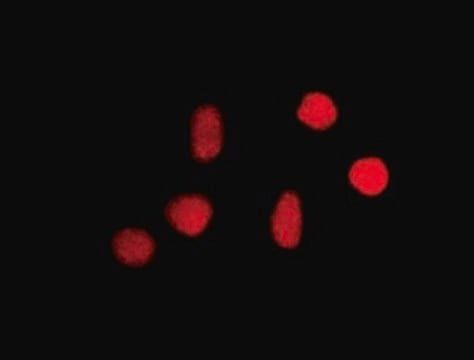
Evaluated by Immunocytochemistry in HeLa cells.

Immunocytochemistry Analysis: A 1:50 dilution of this antibody detected 8-Oxoguanine in HeLa cells.

Linkage

Replaces: MAB3560

Physical form

Format: Purified
Purified mouse monoclonal IgMκ antibody in PBS without preservatives (azide free).

Other Notes

Concentration: Please refer to lot specific datasheet.

Not finding the right product?  

Try our Product Selector Tool.

Storage Class Code

12 - Non Combustible Liquids

WGK

WGK 2

Flash Point(F)

Not applicable

Flash Point(C)

Not applicable


Certificates of Analysis (COA)

Search for Certificates of Analysis (COA) by entering the products Lot/Batch Number. Lot and Batch Numbers can be found on a product’s label following the words ‘Lot’ or ‘Batch’.

Already Own This Product?

Find documentation for the products that you have recently purchased in the Document Library.

Visit the Document Library

Immunofluorescent localization of the murine 8-oxoguanine DNA glycosylase (mOGG1) in cells growing under normal and nutrient deprivation conditions.
Conlon, KA; Zharkov, DO; Berrios, M
DNA Repair null
Diwaker Tripathi et al.
Frontiers in plant science, 11, 596-596 (2020-06-09)
Maize shoot development progresses from non-pigmented meristematic cells at the base of the leaf to expanded and non-dividing green cells of the leaf blade. This transition is accompanied by the conversion of promitochondria and proplastids to their mature forms and
Knut H Lauritzen et al.
DNA repair, 10(6), 639-653 (2011-05-10)
Mitochondria are highly dynamic organelles that can be actively transported within the cell to satisfy local requirements. They are vital for providing cellular energy, but are also an important endogenous source of reactive oxygen species. The distribution of mitochondria is
Alexander P Sobinoff et al.
Toxicological sciences : an official journal of the Society of Toxicology, 118(2), 653-666 (2010-09-11)
Mammalian females are born with a finite number of nonrenewing primordial follicles, the majority of which remain in a quiescent state for many years. Because of their nonrenewing nature, these "resting" oocytes are particularly vulnerable to xenobiotic insult, resulting in
M García-Vaquero et al.
Animal : an international journal of animal bioscience, 6(9), 1435-1443 (2012-10-04)
The aim of this paper is to evaluate at a histopathological level the effect of the most commonly used copper (Cu) supplementation (15 mg/kg dry matter (DM)) in the liver of intensively reared beef cattle. This was done by a

Our team of scientists has experience in all areas of research including Life Science, Material Science, Chemical Synthesis, Chromatography, Analytical and many others.

Contact Technical Service