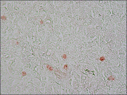
Ki-67 is a nuclear protein, which is expressed in the proliferating cells. Ki-67 is preferentially expressed during late G1-, S-, M-, and G2-phases of the cell cycle, while cells in the G0 (quiescent) phase are negative for this protein.
The gene marker of proliferation Ki-67 (MKI67), spanning 15 exons, is mapped to human chromosome 10q26.2. The encoded protein contains phosphopeptide-binding forkhead-associated (FHA) domain and a protein phosphatase 1 (PP1)-binding site at N- terminal. It possesses 16 tandem repeats at central region and leucine and arginine (LR) pairs at C- terminal end.
Immunogen
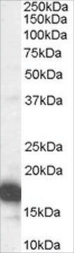
Synthetic peptide from C-terminus of human Ki-67 protein.
Application
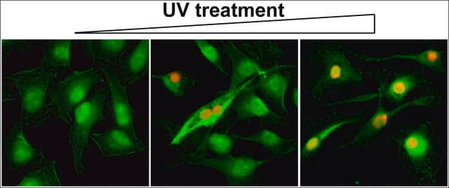
Anti-KI-67 antibody, Rabbit monoclonal has been used in immunohistochemistry.
Biochem/physiol Actions
Marker of proliferation Ki-67 (MKI67), which is a part of the mitotic chromosome periphery, functions as a biological surfactant to maintain individual mitotic chromosomes dispersed in the cytoplasm after nuclear envelope disassembly. Ki-67 is a vital prognostic marker in breast and prostate cancer. Expression of the encoded protein is associated with cell-proliferation rate. Ki-67 is expressed at all active phases of the cell cycle (G (1), S, G (2), and mitosis), except resting cells (G (0)). Therefore, this protein is considered to be a potent cell proliferation marker.
Features and Benefits
Evaluate our antibodies with complete peace of mind. If the antibody does not perform in your application, we will issue a full credit or replacement antibody. Learn more.
Physical form
0.1 ml rabbit monoclonal antibody supplied as tissue culture supernatant in TBS/1% BSA buffer pH 7.5 with less than 0.1% sodium azide.
Disclaimer
Unless otherwise stated in our catalog or other company documentation accompanying the product(s), our products are intended for research use only and are not to be used for any other purpose, which includes but is not limited to, unauthorized commercial uses, in vitro diagnostic uses, ex vivo or in vivo therapeutic uses or any type of consumption or application to humans or animals.
The cancer xenograft model in which human cancer cells are implanted in a mouse is one of the most used preclinical models to test the efficacy of novel cancer drugs. However, the model is imperfect; animal models are ethically burdened
DNA hypermethylation is common in colon cancer. Previously, we have shown that methylation of WNT target genes predicts poor prognosis in stage II colon cancer. The primary objective of this study was to assess whether pre-operative treatment with decitabine can
CD164 regulates proliferation and apoptosis by targeting PTEN in human glioma.
Tu M, et al.
Molecular Medicine Reports, 15(4), 1713-1721 (2017)
The familial dysautonomia disease gene IKBKAP is required in the developing and adult mouse central nervous system.
Proceedings of the National Academy of Sciences of the United States of America, 119(52), e2212306119-e2212306119 (2022-12-20)
Injury to muscle brings about the activation of stem cells, which then generate new myocytes to replace damaged tissue. We demonstrate that this activation is accompanied by a dramatic change in the stem-cell methylation pattern that prepares them epigenetically for
Our team of scientists has experience in all areas of research including Life Science, Material Science, Chemical Synthesis, Chromatography, Analytical and many others.