Skip to Content
Merck
All Photos(3)

Key Documents

SAB4200236

Sigma-Aldrich

Monoclonal Anti-Lamin A/C antibody produced in mouse

clone 4C11, purified from hybridoma cell culture

Synonym(s):

Anti-CDCD1, Anti-CDDC, Anti-CMD1A, Anti-CMT2B1, Anti-EMD2, Anti-FPL, Anti-FPLD, Anti-HGPS, Anti-IDC, Anti-LDP1, Anti-LFP, Anti-LGMD1B, Anti-LMN1, Anti-LMNA, Anti-LMNC, Anti-LMNL1, Anti-PRO1, Anti-renal carcinoma antigen NY-REN-32

Sign Into View Organizational & Contract Pricing


About This Item

MDL number:
UNSPSC Code:
12352203
NACRES:
NA.41

biological source

mouse

conjugate

unconjugated

antibody form

purified immunoglobulin

antibody product type

primary antibodies

clone

4C11, monoclonal

form

buffered aqueous solution

mol wt

antigen ~72/63 kDa

species reactivity

mouse, rat, canine, human, monkey, hamster, bovine

packaging

antibody small pack of 25 μL

concentration

~1.0 mg/mL

technique(s)

immunoprecipitation (IP): suitable
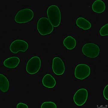
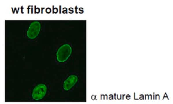
indirect immunofluorescence: suitable
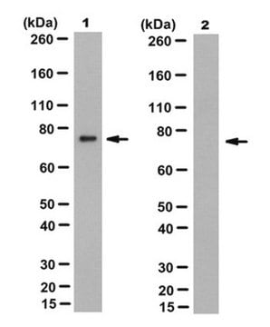
western blot: 0.1-0.2 μg/mL using whole extracts of human HeLa cells

isotype

IgG2a

UniProt accession no.

shipped in

dry ice

storage temp.

−20°C

target post-translational modification

unmodified

Gene Information

human ... LMNA(4000)
mouse ... Lmna(16905)
rat ... Lmna(60374)

General description

Lamin A is a structural protein of the nuclear lamina, a meshwork of intermediate filaments that underlies the inner face of the nuclear envelope. The major components of the nuclear lamina are the lamins that may be classified into two types, A and B. Both A- and B- type lamins are characterized by an a-helical rod domain to enable assembly into filaments, a nuclear localization sequence, and a C-terminal CAAX box isoprenylation sequence for nuclear membrane targeting. A-type lamins, A and C, are produced by alternative splicing resulting in proteins of 664 and 572 amino acids, respectively. The first 566 amino acids of Lamins A and C are identical. Prelamin A, the precursor of Lamin A, has 98 unique amino acids and is farnesylated at its carboxy terminus after synthesis. The last 18 amino acids, which contain the farnesyl group, are removed by an endoproteolytic cleavage, producing the mature Lamin A. Monoclonal Anti-Lamin A/C (mouse IgG2a isotype) is derived from the hybridoma 4C11 produced by the fusion of mouse myeloma cells and splenocytes from BALB/c mice immunized with the Ig-fold domain of human Lamin A.

Specificity

Mouse monoclonal clone 4C11 anti-Lamin A/C antibody recognizes human, rat, mouse, canine, hamster, monkey and bovine Lamin A/C.

Immunogen

hybridoma 4C11 produced by the fusion of mouse myeloma cells and splenocytes from BALB/c mice immunized with the Ig-fold domain of human Lamin A

Application

Mouse monoclonal clone 4C11 anti-Lamin A/C antibody is used to tag lamins A and/or C for detection and quantitation by immunocytochemical and immunohistochemical (IHC) techniques such as immunoblotting, immunoprecipitation, and immunofluorescence. It is used as a probe to determine the presence and roles of lamins A and/or C in nuclear envelope structure and function. This antibody may be used in several immunochemical techniques including immunoblotting (~72/63 kDa), immunoprecipitation and immunofluorescence.

Biochem/physiol Actions

Lamins expressed in somatic cells interact with chromatin, nuclear pore complexes and integral proteins of the inner membrane of the nuclear envelope, such as LAPs 1 and 2 (lamin associated polypeptides), LBR (Lamin B receptor) and emerin. Mutations in Lamin A and C have been linked to a variety of rare human diseases including muscular dystrophy, lipodystrophy, cardiomyopathy, neuropathy and progeroid syndromes (collectively termed laminopathies) and to premature aging (Hutchinson-Gilford progeria syndrome). Lamin A is cleaved into a 47 kDa fragment during apoptosis. Lamin A cleavage seems to be essential for chromatin condensation and nuclear disassembly in apoptosis.

Physical form

Solution in 0.01 M phosphate buffered saline, pH 7.4, containing 15 mM sodium azide

Disclaimer

Unless otherwise stated in our catalog or other company documentation accompanying the product(s), our products are intended for research use only and are not to be used for any other purpose, which includes but is not limited to, unauthorized commercial uses, in vitro diagnostic uses, ex vivo or in vivo therapeutic uses or any type of consumption or application to humans or animals.

Not finding the right product?  

Try our Product Selector Tool.

Storage Class Code

12 - Non Combustible Liquids

WGK

WGK 1

Flash Point(F)

Not applicable

Flash Point(C)

Not applicable


Certificates of Analysis (COA)

Search for Certificates of Analysis (COA) by entering the products Lot/Batch Number. Lot and Batch Numbers can be found on a product’s label following the words ‘Lot’ or ‘Batch’.

Already Own This Product?

Find documentation for the products that you have recently purchased in the Document Library.

Visit the Document Library

M Carolina Gallego Iradi et al.
Scientific reports, 8(1), 4049-4049 (2018-03-08)
To understand how mutations in Matrin 3 (MATR3) cause amyotrophic lateral sclerosis (ALS) and distal myopathy, we used transcriptome and interactome analysis, coupled with microscopy. Over-expression of wild-type (WT) or F115C mutant MATR3 had little impact on gene expression in
The lamin protein family
Dittmer TA and Misteli T
Genome Biology, 12, 222-222 (2011)
Malgorzata Bienkowska-Haba et al.
PLoS pathogens, 14(3), e1006846-e1006846 (2018-03-02)
Herein, we describe a novel infection model that achieves highly efficient infection of primary keratinocytes with human papillomavirus type 16 (HPV16). This cell culture model does not depend on immortalization and is amenable to extensive genetic analyses. In monolayer cell
Nadège Bossuet-Greif et al.
mBio, 9(2) (2018-03-22)
Colibactins are hybrid polyketide-nonribosomal peptides produced by Escherichia coli, Klebsiella pneumoniae, and other Enterobacteriaceae harboring the pks genomic island. These genotoxic metabolites are produced by pks-encoded peptide-polyketide synthases as inactive prodrugs called precolibactins, which are then converted to colibactins by
Veronika Kinterova et al.
Biology of reproduction, 100(4), 896-906 (2018-12-12)
The mechanism of maternal protein degradation during preimplantation development has not been clarified yet. It is thought that a lot of maternal proteins are degraded by the ubiquitin-proteasome system. In this study, we focused on the role of the SCF

Our team of scientists has experience in all areas of research including Life Science, Material Science, Chemical Synthesis, Chromatography, Analytical and many others.

Contact Technical Service