Skip to Content
Merck
All Photos(8)

Key Documents

HPA002643

Sigma-Aldrich

Anti-CASP3 antibody produced in rabbit

enhanced validation

Prestige Antibodies® Powered by Atlas Antibodies, affinity isolated antibody, buffered aqueous glycerol solution

Synonym(s):

Anti-Apopain, Anti-CASP-3, Anti-CPP-32, Anti-Caspase-3 precursor, Anti-Cysteine protease CPP32, Anti-SCA-1, Anti-SREBP cleavage activity 1, Anti-Yama protein

Sign Into View Organizational & Contract Pricing


About This Item

MDL number:
UNSPSC Code:
12352203
Human Protein Atlas Number:
NACRES:
NA.41

biological source

rabbit

Quality Level

conjugate

unconjugated

antibody form

affinity isolated antibody

antibody product type

primary antibodies

clone

polyclonal

product line

Prestige Antibodies® Powered by Atlas Antibodies

form

buffered aqueous glycerol solution

species reactivity

human

enhanced validation

orthogonal RNAseq
Learn more about Antibody Enhanced Validation

technique(s)

immunoblotting: 0.04-0.4 μg/mL
immunofluorescence: 0.25-2 μg/mL
immunohistochemistry: 1:1000-1:2500

immunogen sequence

HGSESMDSGISLDNSYKMDYPEMGLCIIINNKNFHKSTGMTSRSGTDVDAANLRETFRNLKYEVRNKNDLTREEIVELMRDVSKEDHSKRSSFVCVLLSHGEEGIIFGTNGPVDL

UniProt accession no.

shipped in

wet ice

storage temp.

−20°C

target post-translational modification

unmodified

Gene Information

human ... CASP3(836)

General description

CASP3 (caspase 3), the allosteric regulator, is a member of the cysteine-aspartic acid protease (caspase) family which is involved in the inflammation and mammalian apoptosis. It consists of binding sites for small molecules and peptides.

Immunogen

Caspase-3 precursor recombinant protein epitope signature tag (PrEST)

Application

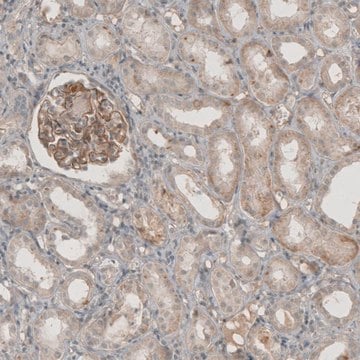
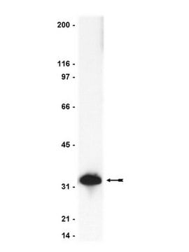
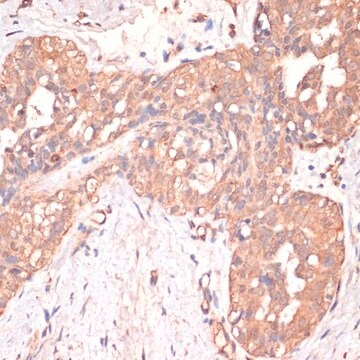
Anti-CASP3 antibody produced in rabbit, a Prestige Antibody, is developed and validated by the Human Protein Atlas (HPA) project . Each antibody is tested by immunohistochemistry against hundreds of normal and disease tissues. These images can be viewed on the Human Protein Atlas (HPA) site by clicking on the Image Gallery link. The antibodies are also tested using immunofluorescence and western blotting. To view these protocols and other useful information about Prestige Antibodies and the HPA, visit sigma.com/prestige.

Biochem/physiol Actions

CASP3 (caspase 3) may have an impact on the melanoma tumor growth after the cytotoxic therapy. It plays a vital role in the proliferation of surrounding cells during executioner phase of apoptosis. Caspases are synthesized and localized as inactive zymogens. After a cascade of proteolytic processing they get activated. Upon activation, they cleave into two separate subunits which further dimerize to form the active enzyme. The activity of caspase-3 is induced by the accumulation of reactive oxygen species (ROS), mitochondrial dysfunction and reduction in adenosine triphosphate (ATP) levels. Alteration in the CASP3 gene is associated with neuronal death in Alzheimer′s disease.

Features and Benefits

Prestige Antibodies® are highly characterized and extensively validated antibodies with the added benefit of all available characterization data for each target being accessible via the Human Protein Atlas portal linked just below the product name at the top of this page. The uniqueness and low cross-reactivity of the Prestige Antibodies® to other proteins are due to a thorough selection of antigen regions, affinity purification, and stringent selection. Prestige antigen controls are available for every corresponding Prestige Antibody and can be found in the linkage section.

Every Prestige Antibody is tested in the following ways:
  • IHC tissue array of 44 normal human tissues and 20 of the most common cancer type tissues.
  • Protein array of 364 human recombinant protein fragments.

Linkage

Corresponding Antigen APREST86566

Physical form

Solution in phosphate-buffered saline, pH 7.2, containing 40% glycerol and 0.02% sodium azide

Legal Information

Prestige Antibodies is a registered trademark of Merck KGaA, Darmstadt, Germany

Disclaimer

Unless otherwise stated in our catalog or other company documentation accompanying the product(s), our products are intended for research use only and are not to be used for any other purpose, which includes but is not limited to, unauthorized commercial uses, in vitro diagnostic uses, ex vivo or in vivo therapeutic uses or any type of consumption or application to humans or animals.

Not finding the right product?  

Try our Product Selector Tool.

Storage Class Code

10 - Combustible liquids

WGK

WGK 1

Flash Point(F)

Not applicable

Flash Point(C)

Not applicable

Personal Protective Equipment

dust mask type N95 (US), Eyeshields, Gloves

Choose from one of the most recent versions:

Certificates of Analysis (COA)

Lot/Batch Number

Don't see the Right Version?

If you require a particular version, you can look up a specific certificate by the Lot or Batch number.

Already Own This Product?

Find documentation for the products that you have recently purchased in the Document Library.

Visit the Document Library

Y Kitamura et al.
Brain research, 780(2), 260-269 (1998-03-21)
Recently, apoptosis has been implicated in the selective neuronal loss of Alzheimer's disease (AD). Apoptosis is regulated by the B cell leukemia-2 gene product (Bcl-2) family (Bcl-2, Bcl-x, Bax, Bak and Bad) and the caspase family (ICH-1 and CPP32), with
Maria Ana Contín et al.
Molecular vision, 19, 1614-1625 (2013-08-01)
Retinal degeneration caused by a defect in the phototransduction cascade leads to the apoptosis of photoreceptor cells, although the precise molecular mechanism is still unknown. In addition, constant low light exposure produces photoreceptor cell death through the activation of downstream
Connexin 43 and Its Hemichannels Mediate Hypoxia?Ischemia-Induced Cell Death in Neonatal Rats
Wang, J
Journal of Child Neurology, 1(1), 1-9 (2014)
Anne L Donato et al.
The Journal of investigative dermatology, 134(6), 1686-1692 (2014-01-18)
Metastatic melanoma often relapses despite cytotoxic treatment, and hence the understanding of melanoma tumor repopulation is crucial for improving our current therapies. In this study, we aim to define the role of caspase 3 in melanoma tumor growth after cytotoxic
Alessandro Rava et al.
Journal of cellular physiology, 237(12), 4563-4579 (2022-11-03)
The loss of NPC1 or NPC2 function results in cholesterol and sphingolipid dyshomeostasis that impairs developmental trajectories, predisposing the postnatal brain to the appearance of pathological signs, including progressive and stereotyped Purkinje cell loss and microgliosis. Despite increasing evidence reporting

Our team of scientists has experience in all areas of research including Life Science, Material Science, Chemical Synthesis, Chromatography, Analytical and many others.

Contact Technical Service