Anti-CD3, mouse monoclonal, clone UCHT1, recognizes the ε-chain of the CD3 complex in human peripheral T cells. It is validated for FC, IP, IHC on frozen sections, and T cell co-stimulation.
Protein A purified mouse monoclonal antibody. Recognizes the ~20 kDa ε chain of the CD3 T-cell receptor complex.
Recognizes the ε-chain of the CD3 complex in human peripheral T cells. Can be used to activate T cells expressing CD3ε.
Immunogen
human thymocytes/Sezary T cells
Application
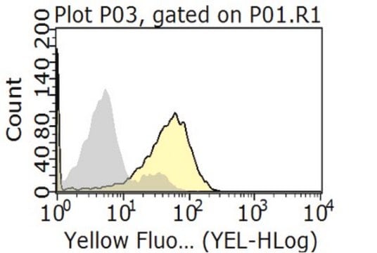
Flow Cytometry (5 µg/ml)
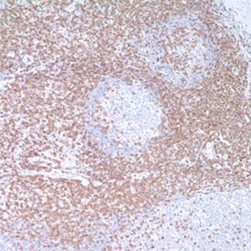
Frozen Sections (see comments)
Immunoprecipitation (see comments)
T cell Co-stimulation (see application references)
Packaging
Please refer to vial label for lot-specific concentration.
Warning
Toxicity: Standard Handling (A)
Other Notes
Thibault, G. and Bardos, P. 1995. J. Immunol.154, 3814. Beverley, P.C., and Callard, R.E. 1981. Eur. J. Immunol.11, 329.
This antibody can be used to activate T cells expressing CD3ε (see application reference). FACS was performed on 5 x 10⁵ cultured HPB-MLT human tumor cells. Optimal binding concentration tested was 5 µg/ml when used with secondary reagent goat anti-mouse IgG, FITC conjugated. This antibody is reported to work for immunoprecipitation and for staining frozen sections. Variables associated with assay conditions will dictate the proper working dilution.
Legal Information
CALBIOCHEM is a registered trademark of Merck KGaA, Darmstadt, Germany
Not finding the right product?
Try our Product Selector Tool.
Storage Class Code
10 - Combustible liquids
WGK
WGK 1
Flash Point(F)
Not applicable
Flash Point(C)
Not applicable
Certificates of Analysis (COA)
Search for Certificates of Analysis (COA) by entering the products Lot/Batch Number. Lot and Batch Numbers can be found on a product’s label following the words ‘Lot’ or ‘Batch’.
Already Own This Product?
Find documentation for the products that you have recently purchased in the Document Library.
American journal of cancer research, 13(11), 5218-5235 (2023-12-07)
This research is dedicated to investigating the mechanism of programmed cell death ligand 1 (PD-L1) and tumor protein 53 target gene 1 (TP53TG1) in immune regulation of colon cancer (CC). Expressions of TP53TG1, PD-L1 and signal transducers and activators of
International journal of biological sciences, 19(16), 5160-5173 (2023-11-06)
Rosacea is a common inflammatory skin disorder mediated by the dysregulation of both keratinocytes and T cells. Here, we report that aquaporin 3 (AQP3), a channel protein that mediates the transport of water/glycerol, was highly expressed in the epidermis and
Frontiers in immunology, 13, 799331-799331 (2022-03-18)
Trace element iron affects T cell biology, but the knowledge about the role of iron in regulating Treg cell expansion is limited. Treg cells play an important role in keeping peripheral T cell tolerance, increasing Treg cell expansion is a
The Journal of clinical investigation, 130(7), 3717-3733 (2020-03-20)
T follicular helper (Tfh) cells are indispensable for the formation of germinal center (GC) reactions, whereas T follicular regulatory (Tfr) cells inhibit Tfh-mediated GC responses. Aberrant activation of Tfh cells contributes substantially to the pathogenesis of autoimmune diseases, such as
The EMBO journal, 40(22), e108125-e108125 (2021-10-08)
Mutations in VAV1, a gene that encodes a multifunctional protein important for lymphocytes, are found at different frequencies in peripheral T-cell lymphoma (PTCL), non-small cell lung cancer, and other tumors. However, their pathobiological significance remains unsettled. After cataloguing 51 cancer-associated
Our team of scientists has experience in all areas of research including Life Science, Material Science, Chemical Synthesis, Chromatography, Analytical and many others.