
HPA004796
Anti-TNFRSF1B antibody produced in rabbit
Prestige Antibodies® Powered by Atlas Antibodies, affinity isolated antibody, buffered aqueous glycerol solution
Synonyme(s) :
Anti-CD120b antigen antibody produced in rabbit, Anti-Etanercept antibody produced in rabbit, Anti-TNF-R2 antibody produced in rabbit, Anti-Tumor necrosis factor receptor 2 antibody produced in rabbit, Anti-Tumor necrosis factor receptor superfamily member 1B precursor antibody produced in rabbit, Anti-Tumor necrosis factor receptor type II antibody produced in rabbit, Anti-p75 antibody produced in rabbit, Anti-p80 TNF-α receptor antibody produced in rabbit
About This Item
Produits recommandés


Source biologique
rabbit
Conjugué
unconjugated
Forme d'anticorps
affinity isolated antibody
Type de produit anticorps
primary antibodies
Clone
polyclonal
Gamme de produits
Prestige Antibodies® Powered by Atlas Antibodies
Forme
buffered aqueous glycerol solution
Espèces réactives
human
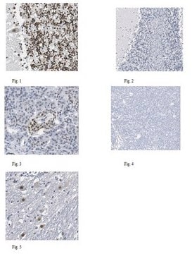
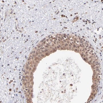
Technique(s)
immunohistochemistry: 1:50-1:200
Séquence immunogène
HAKVFCTKTSDTVCDSCEDSTYTQLWNWVPECLSCGSRCSSDQVETQACTREQNRICTCRPGWYCALSKQEGCRLCAPLRKCRPGFGVARPGTETSDVVCKPCAPGTFSNTTSSTDICRPHQICNVV
Numéro d'accès UniProt
Conditions d'expédition
wet ice
Température de stockage
−20°C
Modification post-traductionnelle de la cible
unmodified
Informations sur le gène
human ... TNFRSF1B(7133)
Description générale
Immunogène
Application
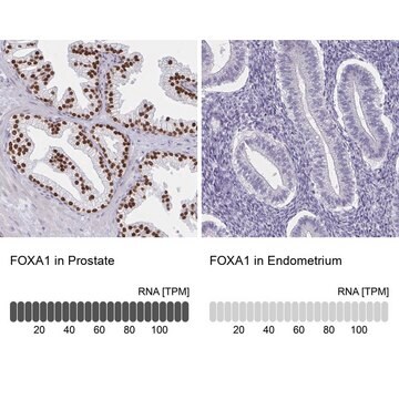
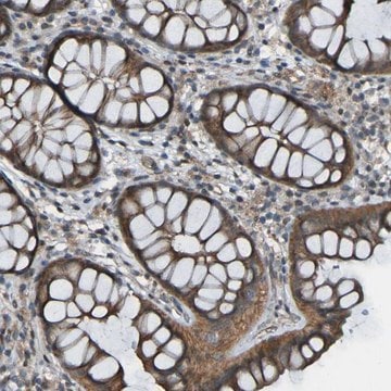
The Human Protein Atlas project can be subdivided into three efforts: Human Tissue Atlas, Cancer Atlas, and Human Cell Atlas. The antibodies that have been generated in support of the Tissue and Cancer Atlas projects have been tested by immunohistochemistry against hundreds of normal and disease tissues and through the recent efforts of the Human Cell Atlas project, many have been characterized by immunofluorescence to map the human proteome not only at the tissue level but now at the subcellular level. These images and the collection of this vast data set can be viewed on the Human Protein Atlas (HPA) site by clicking on the Image Gallery link. We also provide Prestige Antibodies® protocols and other useful information.
Actions biochimiques/physiologiques
Caractéristiques et avantages
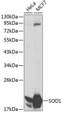
Every Prestige Antibody is tested in the following ways:
- IHC tissue array of 44 normal human tissues and 20 of the most common cancer type tissues.
- Protein array of 364 human recombinant protein fragments.
Liaison
Forme physique
Informations légales
Clause de non-responsabilité
Vous ne trouvez pas le bon produit ?
Essayez notre Outil de sélection de produits.
Code de la classe de stockage
10 - Combustible liquids
Classe de danger pour l'eau (WGK)
WGK 1
Point d'éclair (°F)
Not applicable
Point d'éclair (°C)
Not applicable
Équipement de protection individuelle
Eyeshields, Gloves, multi-purpose combination respirator cartridge (US)
Faites votre choix parmi les versions les plus récentes :
Certificats d'analyse (COA)
Vous ne trouvez pas la bonne version ?
Si vous avez besoin d'une version particulière, vous pouvez rechercher un certificat spécifique par le numéro de lot.
Déjà en possession de ce produit ?
Retrouvez la documentation relative aux produits que vous avez récemment achetés dans la Bibliothèque de documents.
Notre équipe de scientifiques dispose d'une expérience dans tous les secteurs de la recherche, notamment en sciences de la vie, science des matériaux, synthèse chimique, chromatographie, analyse et dans de nombreux autres domaines..
Contacter notre Service technique