Accéder au contenu
Merck
Toutes les photos(1)

Key Documents

MABF2112

Sigma-Aldrich

Anti-EBOV GP Antibody, clone 2G4

clone 2G4, from mouse

Synonyme(s) :

Envelope glycoprotein, GP1, 2, GP

Se connecterpour consulter vos tarifs contractuels et ceux de votre entreprise/organisme


About This Item

Code UNSPSC :
12352203
eCl@ss :
32160702
Nomenclature NACRES :
NA.41

Source biologique

mouse

Forme d'anticorps

purified immunoglobulin

Type de produit anticorps

primary antibodies

Clone

2G4, monoclonal

Espèces réactives

virus, Ebola virus

Conditionnement

antibody small pack of 25 μg

Technique(s)

ELISA: suitable
immunocytochemistry: suitable
immunoprecipitation (IP): suitable
neutralization: suitable

Isotype

IgG2bκ

Numéro d'accès NCBI

Numéro d'accès UniProt

Modification post-traductionnelle de la cible

unmodified

Description générale

Envelope glycoprotein (UniProt: Q05320; also known as GP1,2; GP) is encoded by the GP gene (Gene ID: 911829) in Zaire Ebola virus. Ebola virus (EBOV) is a filovirus that is shown to cause severe viral hemorrhagic fever with high lethality. It has a single stranded, negative-sense RNA genome that encodes seven viral structural proteins including nucleoprotein, virion proteins, and glycoprotein. The glycoprotein is the most important protein involved in pathogenesis. It is a type I transmembrane surface protein shown to be responsible for receptor binding, viral entry, and cellular tropism. The GP gene is reported to undergo transcriptional editing to give rise to several glycosylated proteins, including the structural protein GP1,2, and the secreted non-structural glycoprotein (sGP). The GP is synthesized as a 676-amino acid polyprotein with a signal peptide (aa 1-32). This polyprotein is cleaved by the furin to yield two disulfide linked subunits known as GP1 (aa 33-501) and GP2 (aa 502-676), which together form the GP1,2 heterodimer. The GP1 acts as the receptor-binding subunit and GP2 as the membrane fusion subunit. The GP1 subunit is expressed on the cell surface with the C-terminus oriented toward the aqueous environment, while the N-terminus is bound to GP2 via disulfide bonds. The GP1 subunit contains the heavily glycosylated mucin-like domain (aa 305-485), which is responsible for its cytotoxic function. The glycoprotein contains two coiled coil regions (aa 554-595 and 615-634) that play a role in its oligomerization and fusion activity. Clone 2G4 recognizes glycoprotein lacking a large part of the mucin domain (GP1,2 Mucin333 458). It does not recognize VSV G/ZEBOVGP and subfragment D. When administered before virus challenge, it provides only a partial protection. (Ref.: Qiu, X., et al. (2011). Clin. Immunol. 141(2); 218-227).

Spécificité

Clone 2G4 s a mouse monoclonal antibody that detects envelope glycoprotein of Zaire Ebola virus. It binds to the GP2 region.

Immunogène

Glycoprotein from Zaire Ebola virus, strain Mayinga

Application

Anti-EBOV GP, clone 2G4, Cat. No. MABF2112, is a mouse monoclonal antibody that detects Ebola virus Envelope glycoprotein and has been tested for use in ELISA, Immunocytochemistry, Immunoprecipitation, and Neutralizing Applications.
Immunoprecipitation Analysis: Immunoprecipitation Analysis: A representative lot detected EBOV GP in Immunoprecipitation applications (Qiu, X., et. al. (2011). Clin Immunol. 141(2):218-27).

Immunocytochemistry Analysis: Immunocytochemistry Analysis: A representative lot detected EBOV GP in Immunocytochemistry applications (Qiu, X., et. al. (2011). Clin Immunol. 141(2):218-27).

Neutralizing Analysis: A representative lot provided partial protection when administed prior to virus challenge. (Qiu, X., et. al. (2011). Clin Immunol. 141(2):218-27).

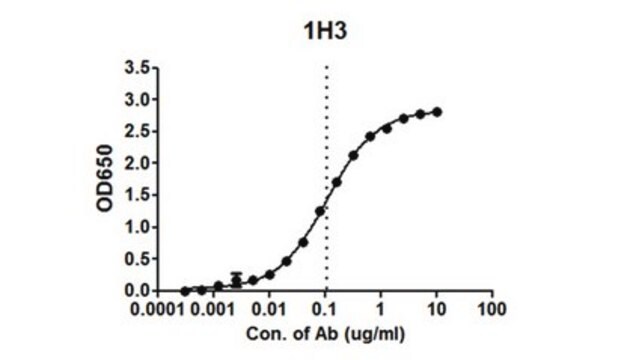
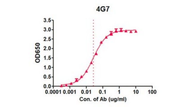
ELISA Analysis: Various dilutions of this antibody detected Envelope glycoprotein in Recombinant Ebola virus Glycoprotein minus the transmembrane region (EBOV rGP TM).

ELISA Analysis: A representative lot detected EBOV GP in ELISA applications (Qiu, X., et. al. (2011). Clin Immunol. 141(2):218-27).
Research Category
Inflammation & Immunology

Qualité

Evaluated by ELISA in Recombinant Ebola virus Glycoprotein minus the transmembrane region (EBOV rGP TM).

ELISA Analysis: Various dilutions of this antibody detected EBOV GP in Recombinant Ebola virus Glycoprotein minus the transmembrane region (EBOV rGP TM).

Description de la cible

74.46 kDa calculated.

Forme physique

Format: Purified
Protein G purified
Purified mouse monoclonal antibody IgG2b in PBS without azide.

Stockage et stabilité

Stable for 1 year at -20°C from date of receipt. Handling Recommendations: Upon receipt and prior to removing the cap, centrifuge the vial and gently mix the solution. Aliquot into microcentrifuge tubes and store at -20°C. Avoid repeated freeze/thaw cycles, which may damage IgG and affect product performance.

Autres remarques

Concentration: Please refer to lot specific datasheet.

Clause de non-responsabilité

Unless otherwise stated in our catalog or other company documentation accompanying the product(s), our products are intended for research use only and are not to be used for any other purpose, which includes but is not limited to, unauthorized commercial uses, in vitro diagnostic uses, ex vivo or in vivo therapeutic uses or any type of consumption or application to humans or animals.

Vous ne trouvez pas le bon produit ?  

Essayez notre Outil de sélection de produits.


Certificats d'analyse (COA)

Recherchez un Certificats d'analyse (COA) en saisissant le numéro de lot du produit. Les numéros de lot figurent sur l'étiquette du produit après les mots "Lot" ou "Batch".

Déjà en possession de ce produit ?

Retrouvez la documentation relative aux produits que vous avez récemment achetés dans la Bibliothèque de documents.

Consulter la Bibliothèque de documents

Notre équipe de scientifiques dispose d'une expérience dans tous les secteurs de la recherche, notamment en sciences de la vie, science des matériaux, synthèse chimique, chromatographie, analyse et dans de nombreux autres domaines..

Contacter notre Service technique