MAB2300
Milli-Mark® Pan Neuronal Marker
Milli-Mark Pan Neuronal Marker is an antibody targeting the Pan Neuronal Marker protein, validated for use in ICC, IHC, IF & IHC.
Synonym(s):
Neuronal Marker
Sign Into View Organizational & Contract Pricing
All Photos(7)
About This Item
UNSPSC Code:
12352203
eCl@ss:
32160702
NACRES:
NA.41
Recommended Products
biological source
mouse
Quality Level
antibody form
purified antibody
clone
monoclonal
species reactivity
rat, mouse, human
manufacturer/tradename
Neuro-Chrom
technique(s)
immunocytochemistry: suitable
immunofluorescence: suitable
immunohistochemistry (formalin-fixed, paraffin-embedded sections): suitable
isotype
IgG1
shipped in
wet ice
target post-translational modification
unmodified
General description
Antibodies to neuronal proteins have become critical tools for identifying neurons and discerning morphological characteristics in culture and complex tissue. While the labeling from classic histological techniques such as Golgi staining and modern molecular approaches such as GFP constructs yield excellent cytoarchitectural detail, these approaches are technically challenging and impractical for many neuroscience research needs. Neuron-specific antibodies are convenient precision tools useful in revealing cytoarchitecture, but are limited to the protein target distribution within the neuron, which may differ greatly from nucleus to soma to dendrite and axon. To achieve as complete a morphological staining as possible across all parts of neurons, Millipore has developed a monoclonal antibody blend that reacts against key somatic, nuclear, dendritic, and axonal proteins distributed across the pan-neuronal architecture that can then be detected by a single secondary antibody. This antibody cocktail has been validated in diverse fixations, cell culture and immunohistochemistry protocols, including archival brain tissue, giving researchers a convienent and specific qualitative and quantitative tool for studying neuronal morphology.
Specificity
Cat. # MAB2300 is specific to axons (neurites), dendrites, nucleus, and the cell body of neurons.
Reactivity with other species has not been determined.
Immunogen
Epitope: Whole Neuron Marker
Application
Research Category
Neuroscience
Neuroscience
Research Sub Category
Neuronal & Glial Markers
Neuronal & Glial Markers
Quality
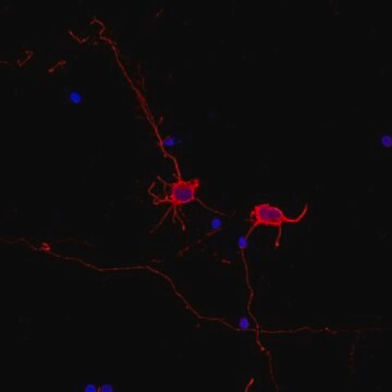
Routinely tested Rat E18 cortex primary neurons in Immunocytochemistry.
Physical form
Blended monoclonal antibody cocktail in PBS with 0.05% NaN3
Format: Purified
Storage and Stability
Maintain at 2-8°C for up to 1 year from date of receipt.
Analysis Note
Control
Rat E18 cortex primary neurons
Rat E18 cortex primary neurons
Legal Information
MILLI-MARK is a registered trademark of Merck KGaA, Darmstadt, Germany
Disclaimer
Unless otherwise stated in our catalog or other company documentation accompanying the product(s), our products are intended for research use only and are not to be used for any other purpose, which includes but is not limited to, unauthorized commercial uses, in vitro diagnostic uses, ex vivo or in vivo therapeutic uses or any type of consumption or application to humans or animals.
Storage Class Code
10 - Combustible liquids
WGK
WGK 2
Flash Point(F)
Not applicable
Flash Point(C)
Not applicable
Certificates of Analysis (COA)
Search for Certificates of Analysis (COA) by entering the products Lot/Batch Number. Lot and Batch Numbers can be found on a product’s label following the words ‘Lot’ or ‘Batch’.
Already Own This Product?
Find documentation for the products that you have recently purchased in the Document Library.
Megan N Evilsizor et al.
Journal of visualized experiments : JoVE, (99), e52293-e52293 (2015-05-21)
Immunohistochemistry is a widely used technique for detecting the presence, location, and relative abundance of antigens in situ. This introductory level protocol describes the reagents, equipment, and techniques required to complete immunohistochemical staining of rodent brain tissue, using markers for
Erdinç Dursun et al.
PloS one, 12(11), e0188605-e0188605 (2017-11-28)
Our recent study indicated that vitamin D and its receptors are important parts of the amyloid processing pathway in neurons. Yet the role of vitamin D receptor (VDR) in amyloid pathogenesis is complex and all regulations over the production of
Harald Lund et al.
Nature immunology, 19(5), 1-7 (2018-04-18)
The cytokine transforming growth factor-β (TGF-β) regulates the development and homeostasis of several tissue-resident macrophage populations, including microglia. TGF-β is not critical for microglia survival but is required for the maintenance of the microglia-specific homeostatic gene signature1,2. Under defined host
Short-term fasting induces profound neuronal autophagy.
Alirezaei, M; Kemball, CC; Flynn, CT; Wood, MR; Whitton, JL; Kiosses, WB
Autophagy null
Muhittin Emre Altunrende et al.
Noro psikiyatri arsivi, 55(4), 295-300 (2019-01-10)
Calcium (Ca) is the phenomenon intracellular molecule that regulate many cellular process in neurons physiologically. Calcium dysregulation may occur in neurons due to excessive synaptic release of glutamate or other reasons related with neurodegeneration. Astaxanthin is a carotenoid that has
Our team of scientists has experience in all areas of research including Life Science, Material Science, Chemical Synthesis, Chromatography, Analytical and many others.
Contact Technical Service


