Skip to Content
Merck
All Photos(1)

Key Documents

MABT336

Sigma-Aldrich

Anti-TKS5 Antibody, clone 13H6.3

clone 13H6.3, from mouse

Synonym(s):

Adapter protein TKS5, Five SH3 domain-containing protein, SH3 multiple domains protein 1, Tyrosine kinase substrate with five SH3 domains, SH3 and PX domain-containing protein 2A

Sign Into View Organizational & Contract Pricing


About This Item

UNSPSC Code:
12352203
eCl@ss:
32160702
NACRES:
NA.41

biological source

mouse

Quality Level

antibody form

purified antibody

antibody product type

primary antibodies

clone

13H6.3, monoclonal

species reactivity

human

packaging

antibody small pack of 25 μg

technique(s)

immunohistochemistry: suitable (paraffin)
western blot: suitable

isotype

IgG1κ

NCBI accession no.

UniProt accession no.

shipped in

ambient

target post-translational modification

unmodified

Gene Information

human ... SH3PXD2A(9644)

General description

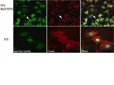
SH3 and PX domain-containing protein 2A (UniProt: Q5TCZ1; also known as Adapter protein TKS5, Five SH3 domain-containing protein, SH3 multiple domains protein 1, Tyrosine kinase substrate with five SH3 domains) is encoded by the SH3PXD2A (also known as FISH, KIAA0418, SH3MD1, TKS5) gene (Gene ID: 9644) in human. Adapter protein TKS5 is a scaffolding protein that is required for podosome formation, degradation of the extracellular matrix, and for invasion in some cancer cells. Three isoforms of TKS5 have been described that are produced by alternative splicing. TKS5 is a cytoplasmic protein in normal cells, but it localizes to podosomes in SRC-transformed cells. It is found in several cancer cell lines, particularly invasive breast carcinomas and melanomas. TKS5 contains one PX (phx homology) domain and five SH3 domains. The PX domain is required for podosome localization because of its ability to bind phosphoinosiltols, such as PtdIns3P and PtdIns(3,4)P2 and to lesser extent PtdIns5P and PtdIns(3,5)P2. Its binding with ADAM12, ADAM15 and ADAM19 is mediated by the fifth SH3 domain. The intramolecular interaction of the PX domain with the third SH3 domain maintains the protein in the cytoplasm and phosphorylation disrupts this interaction, resulting in the redistribution of the protein from cytoplasm to the perimembrane region.

Specificity

Clone 13H6.3 detects SH3 and PX domain-containing protein 2A (adapter protein TKS5) in human cells. It targets and epitope with in 60 amino acids crom the C-terminal half.

Immunogen

GST/His-tagged recombinant fragment corresponding to 60 amino acids from the C-terminal half of human SH3 and PX domain-containing protein 2A (Adapter protein TKS5).

Application

Anti-TKS5, clone 13H6.3, Cat. No. MABT336, ia a mouse monoclonal antibody that detects TKS5 and has been tested for use in Immunohistochemistry (Paraffin) and Western Blotting.
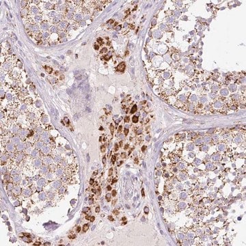
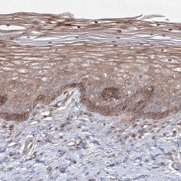
Immunohistochemistry Analysis: A 1:50 dilution from a representative lot detected TKS5 in human breast cancer tissue.
Research Category
Cell Structure

Quality

Evaluated by Western Blotting in SCC61 and SK-MEL28 cell lysates.

Western Blotting Analysis: 0.5 µg/mL of this antibody detected TKS5 in 10 µg of SCC61 and SK-MEL28 cell lysates.

Target description

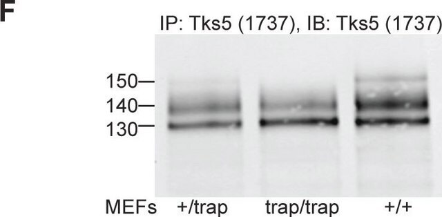
~160 kDa observed; 125.29 kda calculated. Uncharacterized bands may be observed in some lysate(s).

Physical form

Format: Purified
Protein G purified
Purified mouse monoclonal antibody IgG1 in buffer containing 0.1 M Tris-Glycine (pH 7.4), 150 mM NaCl with 0.05% sodium azide.

Storage and Stability

Stable for 1 year at 2-8°C from date of receipt.

Other Notes

Concentration: Please refer to lot specific datasheet.

Disclaimer

Unless otherwise stated in our catalog or other company documentation accompanying the product(s), our products are intended for research use only and are not to be used for any other purpose, which includes but is not limited to, unauthorized commercial uses, in vitro diagnostic uses, ex vivo or in vivo therapeutic uses or any type of consumption or application to humans or animals.

Not finding the right product?  

Try our Product Selector Tool.

Storage Class Code

12 - Non Combustible Liquids

WGK

WGK 1

Flash Point(F)

Not applicable

Flash Point(C)

Not applicable


Certificates of Analysis (COA)

Search for Certificates of Analysis (COA) by entering the products Lot/Batch Number. Lot and Batch Numbers can be found on a product’s label following the words ‘Lot’ or ‘Batch’.

Already Own This Product?

Find documentation for the products that you have recently purchased in the Document Library.

Visit the Document Library

Matthew Baik et al.
MethodsX, 6, 718-726 (2019-04-24)
Invadopodia, cancer cell protrusive structures with associated proteolytic activity, provide cancer cells with the ability to remodel the extracellular matrix. Invadopodia facilitate invasive migration and their formation correlates with cancer cell invasiveness and metastatic potential. The unambiguous identification of invadopodia
Louisiane Perrin et al.
Communications biology, 5(1), 758-758 (2022-08-02)
Invasive and non-invasive cancer cells can invade together during cooperative invasion. However, the events leading to it, role of the epithelial-mesenchymal transition and the consequences this may have on metastasis are unknown. In this study, we demonstrate that the isogenic
Battuya Bayarmagnai et al.
Journal of cell science, 132(20) (2019-09-20)
The process of tumor cell invasion and metastasis includes assembly of invadopodia, protrusions capable of degrading the extracellular matrix (ECM). The effect of cell cycle progression on invadopodia has not been elucidated. In this study, by using invadopodia and cell
Chenxi Tian et al.
Cancer research, 80(7), 1461-1474 (2020-02-08)
The prognosis for pancreatic ductal adenocarcinoma (PDAC) remains poor despite decades of effort. The abundant extracellular matrix (ECM) in PDAC comprises a major fraction of the tumor mass and plays various roles in promoting resistance to therapies. However, nonselective depletion
Juan Liu et al.
Nature metabolism, 4(12), 1830-1846 (2022-12-20)
The glycolytic enzyme lactate dehydrogenase A (LDHA) is frequently overexpressed in cancer, which promotes glycolysis and cancer. The oncogenic effect of LDHA has been attributed to its glycolytic enzyme activity. Here we report an unexpected noncanonical oncogenic mechanism of LDHA;

Our team of scientists has experience in all areas of research including Life Science, Material Science, Chemical Synthesis, Chromatography, Analytical and many others.

Contact Technical Service