Skip to Content
Merck
All Photos(1)

Key Documents

MAB5256X

Sigma-Aldrich

Anti-Neurofilament H Antibody, clone NE14, Alexa Fluor 488 Conjugated

clone NE14, from mouse, ALEXA FLUOR 488

Synonym(s):

200 kDa neurofilament protein, Neurofilament triplet H protein, neurofilament, heavy polypeptide, neurofilament, heavy polypeptide 200kDa

Sign Into View Organizational & Contract Pricing


About This Item

UNSPSC Code:
12352203
eCl@ss:
32160702
NACRES:
NA.41
conjugate:
ALEXA FLUOR 488
application:
ICC
IHC
clone:
NE14, monoclonal
species reactivity:
human, rat, pig, porcine
citations:
5
technique(s):
immunocytochemistry: suitable
immunohistochemistry: suitable

biological source

mouse

Quality Level

conjugate

ALEXA FLUOR 488

antibody form

purified immunoglobulin

antibody product type

primary antibodies

clone

NE14, monoclonal

species reactivity

human, rat, pig, porcine

technique(s)

immunocytochemistry: suitable
immunohistochemistry: suitable

isotype

IgG1

NCBI accession no.

UniProt accession no.

shipped in

wet ice

target post-translational modification

unmodified

Gene Information

human ... NEFH(4744)
pig ... Nefh(100156492)
rat ... Nefh(24587)

General description

Neurofilaments are a type of intermediate filament that serve as major elements of the cytoskeleton supporting the axon cytoplasm. They are the most abundant fibrillar components of the axon, being on average 3-10 times more frequent than axonal microtubules. Neurofilaments (10nm in dia.) are built from three intertwined protofibrils which are themselves composed of two tetrameric protofilament complexes of monomeric proteins. The neurofilament triplet proteins (68/70, 160, and 200 kDa) occur in both the central and peripheral nervous system and are usually neuron specific. The 68/70 kDa NF-L protein can self-assemble into a filamentous structure; however the 160 kDa NF-M and 200 kDa NF-H proteins require the presence of the 68/70 kDa NF-L protein to co-assemble. Neuromas, ganglioneuromas, gangliogliomas, ganglioneuroblastomas and neuroblastomas stain positively for neurofilaments. Although typically restricted to neurons, neurofilaments have been detected in paragangliomas and adrenal and extra-adrenal pheochromocytomas. Carcinoids, neuroendocrine carcinomas of the skin, and oat cell carcinomas of the lung also express neurofilaments.

Specificity

Reactivity with other species has not been tested.
This antibody recognizes Neurofilament H.

Immunogen

Epitope: Unknown
Purified neurofilament polypeptides (Debus et al., 1983).

Application

Anti-Neurofilament H Antibody, clone NE14, Alexa Fluor 488 Conjugated is an antibody against Neurofilament H for use in IH & IC.
Immunohistochemistry:
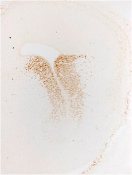
5-10 µg/mL on frozen brain tissue. Optimal working dilutions must be determined by end user.
Research Category
Neuroscience
Research Sub Category
Neurofilament & Neuron Metabolism

Quality

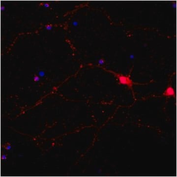
Immunocytochemical Analysis: 1:250 dilution of this lot detected Neurofilament-H in rat cortex cell culture.

Target description

~ 200 kDa

Physical form

Protein G Purified
Purified immunoglobulin in 0.02M Phosphate buffer, 0.25M NaCl containing 0.1% sodium azide.

Storage and Stability

Stable for 6 months at 2-8ºC from date of receipt.

Analysis Note

Control
Rat Cortex Cells.

Other Notes

Concentration: Please refer to the Certificate of Analysis for the lot-specific concentration.

Legal Information

ALEXA FLUOR is a trademark of Life Technologies

Disclaimer

Unless otherwise stated in our catalog or other company documentation accompanying the product(s), our products are intended for research use only and are not to be used for any other purpose, which includes but is not limited to, unauthorized commercial uses, in vitro diagnostic uses, ex vivo or in vivo therapeutic uses or any type of consumption or application to humans or animals.

Not finding the right product?  

Try our Product Selector Tool.

Storage Class Code

12 - Non Combustible Liquids

WGK

WGK 2

Flash Point(F)

Not applicable

Flash Point(C)

Not applicable


Certificates of Analysis (COA)

Search for Certificates of Analysis (COA) by entering the products Lot/Batch Number. Lot and Batch Numbers can be found on a product’s label following the words ‘Lot’ or ‘Batch’.

Already Own This Product?

Find documentation for the products that you have recently purchased in the Document Library.

Visit the Document Library

Emiri Miura-Yura et al.
Journal of diabetes investigation, 11(1), 28-38 (2019-05-31)
Transplantation of stem cells promotes axonal regeneration and angiogenesis in a paracrine manner. In the present study, we examined whether the secreted factors in conditioned medium of stem cells from human exfoliated deciduous teeth (SHED-CM) had beneficial effects on diabetic
Sienna S Drake et al.
International journal of molecular sciences, 23(23) (2022-12-12)
Multiple sclerosis (MS) is an autoimmune and neurodegenerative disease driven by inflammation and demyelination in the brain, spinal cord, and optic nerve. Optic neuritis, characterized by inflammation and demyelination of the optic nerve, is a symptom in many patients with
Sandeep Sheth et al.
Scientific reports, 9(1), 9571-9571 (2019-07-04)
Adenosine A1 receptors (A1AR) are well characterized for their role in cytoprotection. Previous studies have demonstrated the presence of these receptors in the cochlea where their activation were shown to suppress cisplatin-induced inflammatory response and the resulting ototoxicity. Inhibition of
Hiromi Nakai-Shimoda et al.
iScience, 25(1), 103609-103609 (2022-01-11)
Glucose-responsive ATP-sensitive potassium channels (KATP) are expressed in a variety of tissues including nervous systems. The depolarization of the membrane potential induced by glucose may lead to hyperexcitability of neurons and induce excitotoxicity. However, the roles of KATP in the
S S Oh et al.
Neuroscience, 212, 201-213 (2012-04-21)
The contribution of oxidative stress to diabetic complications including neuropathy is widely known. Mitochondrial and cellular damage are associated with the overproduction of reactive oxygen species and decreased levels or function of the cellular antioxidant mitochondrial manganese superoxide dismutase (SOD2).

Our team of scientists has experience in all areas of research including Life Science, Material Science, Chemical Synthesis, Chromatography, Analytical and many others.

Contact Technical Service