Skip to Content
Merck
All Photos(1)

Key Documents

396M-1

Sigma-Aldrich

Parvalbumin (2E11) Mouse Monoclonal Antibody

Sign Into View Organizational & Contract Pricing


About This Item

UNSPSC Code:
12352202
NACRES:
NA.41

biological source

mouse

conjugate

unconjugated

antibody form

culture supernatant

antibody product type

primary antibodies

clone

2E11, monoclonal

description

For In Vitro Diagnostic Use in Select Regions (See Chart)

form

buffered aqueous solution

species reactivity

human

packaging

vial of 0.1 mL concentrate (396M-14)
vial of 0.5 mL concentrate (396M-15)
bottle of 1.0 mL predilute (396M-17)
vial of 1.0 mL concentrate (396M-16)
bottle of 7.0 mL predilute (396M-18)

manufacturer/tradename

Cell Marque

technique(s)

immunohistochemistry (formalin-fixed, paraffin-embedded sections): 1:10-1:50

isotype

IgG1

control

chromophobe renal cell carcinoma

shipped in

wet ice

storage temp.

2-8°C

visualization

cytoplasmic

Gene Information

human ... PVALB(5816)

General description

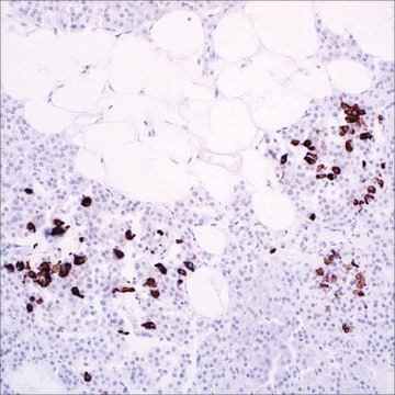
Parvalbumin is a calcium-binding protein that regulates cytosolic calcium homeostasis. Tissue types that express this protein have been reported in the muscle, brain, neuroendocrine organs, and kidney.In normal kidney, parvalbumin has been shown to be limited to the distal tubular and collecting duct cells (the intercalated cells). Parvalbumin is strongly expressed in almost all primary, as well as metastatic, chromophobe renal cell carcinoma (RCC) (100%) and oncocytoma (69%), but is essentially negative in other types of RCCs, such as clear cell RCC and papillary RCC. This limited expression has been considered in keeping with the putative histogenesis of chromophobe RCC and oncocytoma from the distal portion of the nephron and may facilitate in their differential diagnoses.

Quality


IVD

IVD

IVD

RUO

Linkage

Parvalbumin Positive Control Slides, Product No. 396S, are available for immunohistochemistry (formalin-fixed, paraffin-embedded sections).

Physical form

Solution in Tris Buffer, pH 7.3-7.7, with 1% BSA and <0.1% Sodium Azide

Preparation Note

Download the IFU specific to your product lot and formatNote: This requires a keycode which can be found on your packaging or product label.

Other Notes

For Technical Service please contact: 800-665-7284 or email: service@cellmarque.com

Legal Information

Cell Marque is a trademark of Merck KGaA, Darmstadt, Germany

Not finding the right product?  

Try our Product Selector Tool.


Certificates of Analysis (COA)

Search for Certificates of Analysis (COA) by entering the products Lot/Batch Number. Lot and Batch Numbers can be found on a product’s label following the words ‘Lot’ or ‘Batch’.

Already Own This Product?

Find documentation for the products that you have recently purchased in the Document Library.

Visit the Document Library

Brian P Adley et al.
Analytical and quantitative cytology and histology, 28(4), 228-236 (2006-08-25)
To investigate the diagnostic value of cytokeratin 7 (CK7) and parvalbumin at mRNA and protein levels. CK7 and parvalbumin mRNA expression levels in 23 oncocytomas and 32 chromophobe renal cell carcinomas (RCCs) were examined using gene expression microarrays. Immunohistochemistry was
Pusiol Teresa et al.
Diagnostic cytopathology, 40(1), 56-61 (2011-12-20)
Malignant effusions because of renal-cell carcinoma (RCC) are an unusual event and occur in patients with papillary and clear cell tumors. We have studied a 65-year-old man who underwent right renal tumorectomy, diagnosed as chromophobe RCC (pT1). After 16 months
G Martignoni et al.
Modern pathology : an official journal of the United States and Canadian Academy of Pathology, Inc, 14(8), 760-767 (2001-08-16)
Chromophobe renal carcinoma is composed of neoplastic cell showing several features similar to those found in the intercalated cells of the collecting ducts. Because the distal nephron expresses calcium-binding proteins playing a role in calcium homeostasis, we reasoned that these
Andrew N Young et al.
The American journal of surgical pathology, 27(2), 199-205 (2003-01-28)
The common histopathologic subtypes of renal epithelial neoplasms include conventional, or clear cell, renal cell carcinoma (RCC), papillary RCC, chromophobe RCC, and renal oncocytoma. These subtypes differ clinically and pathologically, making accurate classification important. However, this differential diagnosis can be

Our team of scientists has experience in all areas of research including Life Science, Material Science, Chemical Synthesis, Chromatography, Analytical and many others.

Contact Technical Service